

Comparison of cilomilast, tadalafil, and both drug combinations in the treatment of monocrotaline-induced pulmonary arterial hypertension in rats
- PMID: 40783516
- PMCID: PMC12335037
- DOI: 10.1186/s12872-025-05065-0
Comparison of cilomilast, tadalafil, and both drug combinations in the treatment of monocrotaline-induced pulmonary arterial hypertension in rats
Abstract
Background: Pulmonary arterial hypertension (PAH) is a progressive disease characterized by endothelial dysfunction and inflammation. This study aimed to evaluate the effects of cilomilast (CIL), a phosphodiesterase-4 inhibitor, and tadalafil (TAD), a phosphodiesterase-5 inhibitor, on PAH induced by monocrotaline (MCT) in rats.
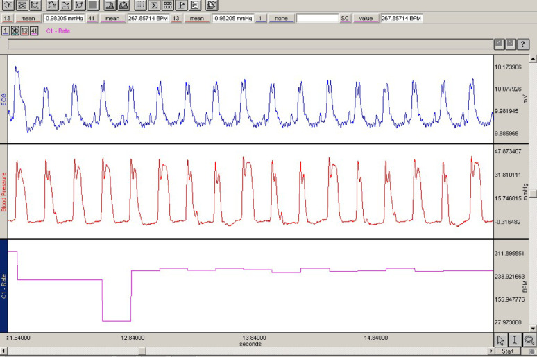
Methods: Forty Wistar albino rats were divided into five groups: control, MCT, MCT + CIL, MCT + TAD, and MCT + CIL + TAD. PAH was induced via MCT, and treatments were administered orally from days 21 to 35. Hemodynamic parameters, right ventricular pressure (RVP), echocardiographic findings, and histopathological lung and heart tissue changes were assessed. Nitric oxide (NO) levels in lung tissue were also measured.
Results: Tissue NO levels were significantly greater in the MCT + CIL + TAD group than in the MCT group (p = 0.01). The RVP was lower in the MCT + TAD and MCT + CIL + TAD groups than in the MCT group (p < 0.05) but not in the MCT + CIL group. Histopathologically, lung perivascular infiltration and pulmonary artery wall thickness were significantly reduced in the MCT + CIL + TAD group, indicating an anti-inflammatory effect. However, CIL alone did not significantly impact pulmonary artery thickening or RVP.
Conclusion: CIL alone had no significant effect on PAH progression, but its combination with TAD improved inflammation scores and NO levels. These findings suggest that targeting inflammation alongside vasodilation may offer therapeutic benefits in PAH. Further studies with different doses and PAH models are recommended.
Keywords: Cilomilast; Monocrotaline; Pulmonary arterial hypertension; Rat; Tadalafil.
© 2025. The Author(s).
Conflict of interest statement
Declarations. Ethics approval and consent to participate: Ethical approval was obtained from the Experimental Animal Ethics Board at İnönü University (2016 A-114). Consent for publication: Not applicable. Competing interests: The authors declare no competing interests.
Figures


Similar articles
-
C-C Motif chemokine receptor-2 blockade ameliorates pulmonary hypertension in rats and synergizes with a pulmonary vasodilator.Cardiovasc Res. 2025 Jul 8;121(7):1076-1090. doi: 10.1093/cvr/cvae244. Cardiovasc Res. 2025. PMID: 39556088 Free PMC article.
-
Sulforaphane Improves Redox Homeostasis and Right Ventricular Contractility in a Model of Pulmonary Hypertension.J Cardiovasc Pharmacol. 2024 Jun 1;83(6):612-620. doi: 10.1097/FJC.0000000000001557. J Cardiovasc Pharmacol. 2024. PMID: 38547510
-
Salidroside ameliorates monocrotaline-induced pulmonary arterial hypertension in rats by modulating BKCa channels.Eur J Pharmacol. 2025 Oct 5;1004:177950. doi: 10.1016/j.ejphar.2025.177950. Epub 2025 Jul 24. Eur J Pharmacol. 2025. PMID: 40714070
-
Systemic treatments for metastatic cutaneous melanoma.Cochrane Database Syst Rev. 2018 Feb 6;2(2):CD011123. doi: 10.1002/14651858.CD011123.pub2. Cochrane Database Syst Rev. 2018. PMID: 29405038 Free PMC article.
-
Current Overview of the Biology and Pharmacology in Sugen/Hypoxia-Induced Pulmonary Hypertension in Rats.J Aerosol Med Pulm Drug Deliv. 2024 Oct;37(5):241-283. doi: 10.1089/jamp.2024.0016. J Aerosol Med Pulm Drug Deliv. 2024. PMID: 39388691 Review.
References
-
- Hautefort A, Girerd B, Montani D, Cohen-Kaminsky S, Price L, Lambrecht BN, Humbert M, Perros F. T-helper 17 cell polarization in pulmonary arterial hypertension. Chest. 2015;147(6):1610–20. - PubMed
Publication types
MeSH terms
Substances
Grants and funding
LinkOut - more resources
Full Text Sources
Medical
