Review
. 2025 Aug 10;150(1):16.
doi: 10.1007/s00401-025-02923-1.

A focus on the normal-appearing white and gray matter within the multiple sclerosis brain: a link to smoldering progression

Affiliations
Review

A focus on the normal-appearing white and gray matter within the multiple sclerosis brain: a link to smoldering progression

Gema Muñoz González et al. Acta Neuropathol. .

Abstract

Multiple sclerosis is a chronic neuro-inflammatory and neurodegenerative disease, traditionally characterized by the presence of focal demyelinating lesions in the CNS. However, accumulating evidence suggests that multiple sclerosis pathophysiology extends beyond such classical lesions, affecting also 'normal' appearing tissue in both white and gray matter, referred to as 'normal-appearing white matter' and 'normal-appearing gray matter', respectively. Here, we provide a comprehensive overview of the widespread biochemical, cellular, and microstructural alterations occurring in these 'normal-appearing' CNS regions. Additionally, we discuss the evidence derived from human post-mortem studies that support that normal-appearing white and gray matter could be the drivers of smoldering-associated pathological worsening once repair mechanisms are exhausted. Comprehensive understanding of multiple sclerosis pathology beyond classical lesions not only provides a more complete picture of disease progression, but also provides further insights into potential novel therapeutic avenues in order to slow or halt disability accumulation.

Keywords: Degeneration; Myelin; Oligodendrocyte; Smoldering disease.


Conflict of interest statement

Declarations. Conflict of interest: The authors declare no competing interests.

Figures

Fig. 1
Fig. 1
Natural clinical course of Multiple Sclerosis (MS). Relapse-associated worsening (RAW) is more frequent during the early phases of MS but decreases as the disease progresses. In contrast, smoldering-associated worsening (SAW) gradually accumulates and becomes more noticeable over time, particularly as relapses diminish. SAW is the primary driver of disability accumulation, even in the early stages of the disease
Fig. 2
Fig. 2
Pathological correlates in Multiple Sclerosis (MS) compared to control CNS tissue. Key pathological changes in MS, including alterations in normal-appearing white matter (NAWM) and normal-appearing grey matter (NAGM), diffusely abnormal white matter (DAWM) and micro-diffusely abnormal white matter (mDAWM), meningeal inflammation-associated damage, atrophy, and MS lesions
Fig. 3
Fig. 3
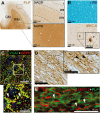
Pathological alterations in normal-appearing gray matter (NAGM) and white matter (NAWM) in Multiple Sclerosis (MS). A Representative image of an MS brain tissue section stained with an antibody against PLP, highlighting regions of both NAGM and NAWM. B Luxol Fast Blue (LFB) staining (top) reveals compact and homogeneous lipid distribution within NAWM. In the same region, staining for MHC-II (bottom) shows microglial clustering. C Staining for vGAT (a marker of inhibitory presynaptic terminals) and Gephyrin (a postsynaptic inhibitory marker). Co-localization (yellow) alternates with areas where only postsynaptic Gephyrin is detected, indicating loss of inhibitory presynaptic terminals. D SMI312 staining in NAWM highlights axonal transections (*) and swellings (arrow bars), reflecting ongoing axonal degeneration. E Dual staining for PLP and SMI312 in NAWM reveals local detachments of the myelin sheath from the underlying axons (“myelin blisters”)
Fig. 4
Fig. 4
Overview of widespread abnormalities in normal-appearing gray matter (NAGM) and normal-appearing white matter (NAWM) that may synergistically contribute to disability accumulation. (1) Metabolic and functional dysregulation of oligodendrocyte progenitor cells, oligodendrocytes, and myelin may result from cell-intrinsic mechanisms, from damage from nearby lesions or diffusely abnormal white matter, or the overall proinflammatory environment within the MS brain. Myelin segments, possibly with aberrant lipid profile and/or excessive MBP citrullination, may have a reduced attachment between the lamella, and therefore may decompact or present with structural abnormalities, such as myelin blisters or myelinosomes. Oligodendrocyte stress and myelin alterations could thus favor demyelination. (2) Microglial nodules form around partially demyelinated axons and actively phagocytose myelin debris. While some nodules likely resolve, others may persist and evolve into chronic microglial activation, adopting a proinflammatory phenotype that impairs the repair process. Myelin damage may be further exacerbated by microglia- and lymphocyte-derived factors, such as proinflammatory cytokines and reactive oxygen species. In the context of a persistently proinflammatory environment, intrinsic oligodendrocyte dysfunction and secondary microglial responses may converge to disrupt the myelin sheath. This cascade can initiate a ripple effect leading to widespread demyelination and lesion formation, potentially amplified by lymphocyte infiltration through blood vessels. (3) Axonal energy deficiency, whether caused by subtle myelin damage or overt demyelination, may elicit adaptive neuronal responses, including an increased number of axonal mitochondria. However, the failure of these compensatory mechanisms—alongside retrograde and Wallerian degeneration originating from distal lesions—ultimately contributes to severe axonal damage. (4) In the NAGM, neuronal stress caused by axonal damage in the NAWM, nearby or distant lesions, and meningeal inflammation can result in subtle signs of neuronal injury—such as synaptic puncta retraction—and may ultimately lead to neuronal loss, particularly in subpopulations with the greatest energetic demands. OPCs  oligodendrocyte progenitor cells, ROS reactive oxygen species, CRYAB   α-crystallin B, JPN  juxtaparanodes, PN   paranodes, NoR  nodes of Ranvier, citMBP  citrullinated myelin basic protein

Similar articles

References

    1. Absinta M, Cortese ICM, Vuolo L, Nair G, de Alwis MP, Ohayon J et al (2017) Leptomeningeal gadolinium enhancement across the spectrum of chronic neuroinflammatory diseases. Neurology 88:1439–1444. 10.1212/WNL.0000000000003820 - PMC - PubMed
    1. Albert M, Barrantes-Freer A, Lohrberg M, Antel JP, Prineas JW, Palkovits M et al (2017) Synaptic pathology in the cerebellar dentate nucleus in chronic multiple sclerosis. Brain Pathol 27:737–747. 10.1111/bpa.12450 - PMC - PubMed
    1. Arancibia-Carcamo IL, Attwell D (2014) The node of ranvier in CNS pathology. Acta Neuropathol 128:161–175. 10.1007/s00401-014-1305-z - PMC - PubMed
    1. Arancibia-Cárcamo IL, Ford MC, Cossell L, Ishida K, Tohyama K, Attwell D (2017) Node of ranvier length as a potential regulator of myelinated axon conduction speed. Elife 6:e23329. 10.7554/eLife.23329 - PMC - PubMed
    1. Asadollahi E, Trevisiol A, Saab AS, Looser ZJ, Dibaj P, Ebrahimi R et al (2024) Oligodendroglial fatty acid metabolism as a central nervous system energy reserve. Nat Neurosci 27:1934–1944. 10.1038/s41593-024-01749-6 - PMC - PubMed

LinkOut - more resources