Mitochondrial DNA methylation is involved in contrast-induced renal tubular epithelial cell injury
- PMID: 40784878
- PMCID: PMC12337735
- DOI: 10.1080/0886022X.2025.2532112
Mitochondrial DNA methylation is involved in contrast-induced renal tubular epithelial cell injury
Abstract
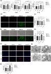
Mitochondrial DNA (mtDNA) methylation may be associated with mitochondrial damage; this study investigates their relationship in contrast-induced renal tubular epithelial cell (RTEC) injury. We stimulated HK-2 cells with iohexol to establish an in vitro model and analyzed the methylation level of mtDNA by bisulfite amplicon sequencing. The mitochondrial membrane potential, mitochondrial reactive oxygen species (mtROS), intracellular ROS, and changes in mitochondrial ultrastructure were evaluated as indicators of mitochondrial damage. Iohexol significantly inhibited cell viability and induced cell apoptosis, increasing both mtROS and intracellular ROS levels. Additionally, the methylation levels of mtDNA-encoded genes cytochrome c oxidase subunit I (COX I) (3.09%, *p < 0.05), cytochrome c oxidase subunit II (COX II) (4.51%, **p < 0.01), cytochrome c oxidase subunit III (COX III) (3.50%, **p < 0.01) and cytochrome B (CYTB)(4.66%, *p < 0.05) were increased, accompanied by enhanced transcription of both COX I and COX III. 5-Aza-dC, as a DNA methylation inhibitor, was dissolved in dimethyl sulfoxide (DMSO) vehicle to explore the role and mechanism of inhibiting mtDNA methylation in contrast-induced RTEC injury. HK-2 cells were further divided into four groups: vehicle control (DMSO alone), vehicle pretreated contrast - induced group (CI) (DMSO-CI), inhibitor control (5-Aza-dC), and inhibitor pretreated CI (5-Aza-dC-CI). Intriguingly, administration of 5-Aza-dC effectively attenuated mtDNA methylation, leading to improvements in these parameters and restoration of cell viability while reducing apoptosis. In conclusion, mtDNA methylation is involved in the mechanism of contrast-induced RTEC injury, potentially mediated by over-transcription of COX I and III, abnormal mtROS production, and subsequent mitochondrial damage and dysfunction. Inhibiting mtDNA methylation can provide protective effects against contrast - induced RTEC injury by reducing ROS (mtROS) production.
Keywords: CI-AKI; Mitochondrial DNA methylation; ROS; apoptosis; mitochondrial damage.
Conflict of interest statement
The authors declare that the research was conducted in the absence of any commercial or financial relationships that could be construed as a potential conflict of interest.
Figures


Similar articles
-
Glycyrrhizin alleviates contrast-induced acute kidney injury via inhibiting HMGB1-mediated renal tubular epithelial cells ferroptosis.Ren Fail. 2025 Dec;47(1):2548613. doi: 10.1080/0886022X.2025.2548613. Epub 2025 Aug 31. Ren Fail. 2025. PMID: 40887103 Free PMC article.
-
Critical roles of tubular mitochondrial ATP synthase dysfunction in maleic acid-induced acute kidney injury.Apoptosis. 2024 Jun;29(5-6):620-634. doi: 10.1007/s10495-023-01897-3. Epub 2024 Jan 28. Apoptosis. 2024. PMID: 38281282 Free PMC article.
-
Mito-tempo ameliorates tubular injury of diabetic nephropathy via inhibiting mt-dsRNA release and PKR/eIF2α pathway activation.Free Radic Biol Med. 2025 Sep;237:147-159. doi: 10.1016/j.freeradbiomed.2025.05.431. Epub 2025 May 31. Free Radic Biol Med. 2025. PMID: 40456498
-
Management of urinary stones by experts in stone disease (ESD 2025).Arch Ital Urol Androl. 2025 Jun 30;97(2):14085. doi: 10.4081/aiua.2025.14085. Epub 2025 Jun 30. Arch Ital Urol Androl. 2025. PMID: 40583613 Review.
-
Regulation of mitochondrial oxidative phosphorylation through tight control of cytochrome c oxidase in health and disease - Implications for ischemia/reperfusion injury, inflammatory diseases, diabetes, and cancer.Redox Biol. 2024 Dec;78:103426. doi: 10.1016/j.redox.2024.103426. Epub 2024 Nov 10. Redox Biol. 2024. PMID: 39566165 Free PMC article. Review.
References
MeSH terms
Substances
LinkOut - more resources
Full Text Sources
