

Immunological synapse: structures, molecular mechanisms and therapeutic implications in disease
- PMID: 40784895
- PMCID: PMC12336355
- DOI: 10.1038/s41392-025-02332-6
Immunological synapse: structures, molecular mechanisms and therapeutic implications in disease
Abstract
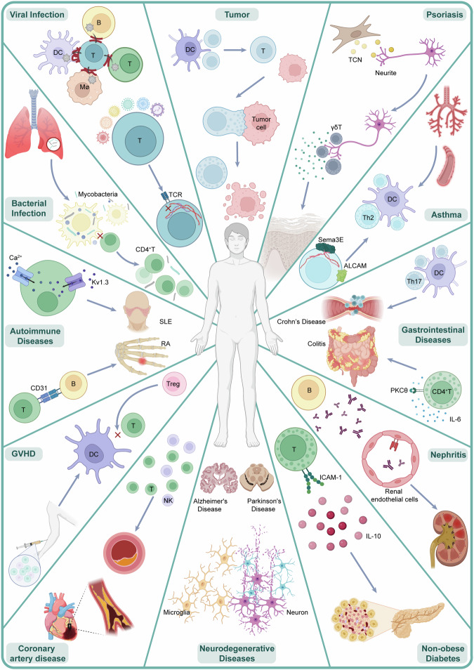
The immunological synapse (IS) serves as the fundamental architectural framework for direct interactions and secretory crosstalk between immune cells, as well as between immune cells and other cells. Its dysregulation is thought to be a key underlying cause of immune evasion or inflammation observed in various diseases, including tumors and infections. Numerous recent studies have addressed key signaling mechanisms and reported novel targets related to IS, further broadening our understanding of its function and regulatory factors. However, a comprehensive review that highlights recent progress and consolidates past knowledge is still lacking. In this study, we delineated the pre- and postsynaptic structures constituting the IS between T cells, natural killer (NK) cells, dendritic cells (DCs), and macrophages. We also detail the specific signaling mechanisms and pathways that modulate the formation and disassembly of the IS, including cytoskeletal remodeling, membrane reshaping, integrin signaling, and force transduction. Following these experimental findings, we systematically review the central roles of IS in maintaining homeostasis and health and outline various diseases arising from IS disorders. Finally, we thoroughly explore targets and treatments related to IS on the basis of preclinical evidence and clinical trials, with the aim of providing further investigatory and therapeutic insights for researchers and clinicians.
© 2025. The Author(s).
Conflict of interest statement
Competing interests: The authors declare no competing interests.
Figures
Similar articles
-
Systemic pharmacological treatments for chronic plaque psoriasis: a network meta-analysis.Cochrane Database Syst Rev. 2020 Jan 9;1(1):CD011535. doi: 10.1002/14651858.CD011535.pub3. Cochrane Database Syst Rev. 2020. Update in: Cochrane Database Syst Rev. 2021 Apr 19;4:CD011535. doi: 10.1002/14651858.CD011535.pub4. PMID: 31917873 Free PMC article. Updated.
-
Systemic pharmacological treatments for chronic plaque psoriasis: a network meta-analysis.Cochrane Database Syst Rev. 2021 Apr 19;4(4):CD011535. doi: 10.1002/14651858.CD011535.pub4. Cochrane Database Syst Rev. 2021. Update in: Cochrane Database Syst Rev. 2022 May 23;5:CD011535. doi: 10.1002/14651858.CD011535.pub5. PMID: 33871055 Free PMC article. Updated.
-
Systemic pharmacological treatments for chronic plaque psoriasis: a network meta-analysis.Cochrane Database Syst Rev. 2017 Dec 22;12(12):CD011535. doi: 10.1002/14651858.CD011535.pub2. Cochrane Database Syst Rev. 2017. Update in: Cochrane Database Syst Rev. 2020 Jan 9;1:CD011535. doi: 10.1002/14651858.CD011535.pub3. PMID: 29271481 Free PMC article. Updated.
-
Short-Term Memory Impairment.2024 Jun 8. In: StatPearls [Internet]. Treasure Island (FL): StatPearls Publishing; 2025 Jan–. 2024 Jun 8. In: StatPearls [Internet]. Treasure Island (FL): StatPearls Publishing; 2025 Jan–. PMID: 31424720 Free Books & Documents.
-
T-bet expressing Tr1 cells driven by dietary signals dominate the small intestinal immune landscape.bioRxiv [Preprint]. 2025 Jul 4:2025.06.30.662190. doi: 10.1101/2025.06.30.662190. bioRxiv. 2025. PMID: 40747421 Free PMC article. Preprint.
References
-
- Harwood, N. E. & Batista, F. D. Early events in B cell activation. Annu. Rev. Immunol.28, 185–210 (2010). - PubMed
-
- Delon, J., Kaibuchi, K. & Germain, R. N. Exclusion of CD43 from the immunological synapse is mediated by phosphorylation-regulated relocation of the cytoskeletal adaptor moesin. Immunity15, 691–701 (2001). - PubMed
-
- Allenspach, E. J. et al. ERM-dependent movement of CD43 defines a novel protein complex distal to the immunological synapse. Immunity15, 739–750 (2001). - PubMed
-
- Lee, K.-H. et al. T cell receptor signaling precedes immunological synapse formation. Science295, 1539–1542 (2002). - PubMed
-
- Al-Alwan, M. M., Rowden, G., Lee, T. D. & West, K. A. The dendritic cell cytoskeleton is critical for the formation of the immunological synapse. J. Immunol.166, 1452–1456 (2001). - PubMed
Publication types
MeSH terms
Grants and funding
LinkOut - more resources
Full Text Sources
Medical
Miscellaneous
