. 2025 Aug 10;15(1):29277.
doi: 10.1038/s41598-025-15563-y.

Harnessing electroacupuncture: a promising strategy against sleep deprivation-exacerbated post-cardiac arrest brain injury

Affiliations

Harnessing electroacupuncture: a promising strategy against sleep deprivation-exacerbated post-cardiac arrest brain injury

Wenchao Fu et al. Sci Rep. .

Abstract

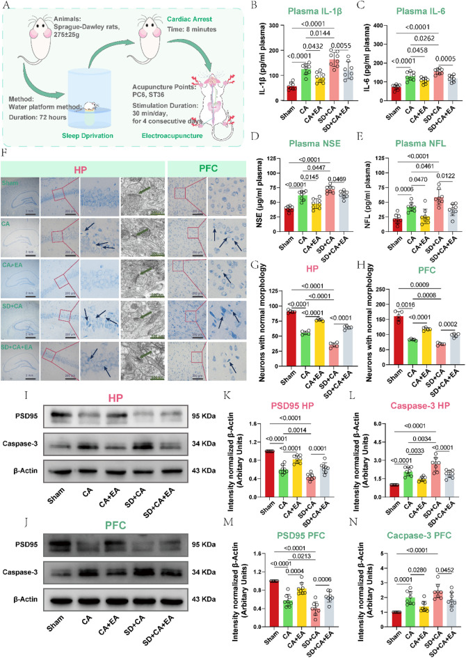
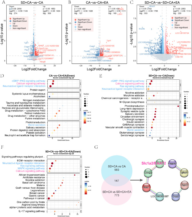
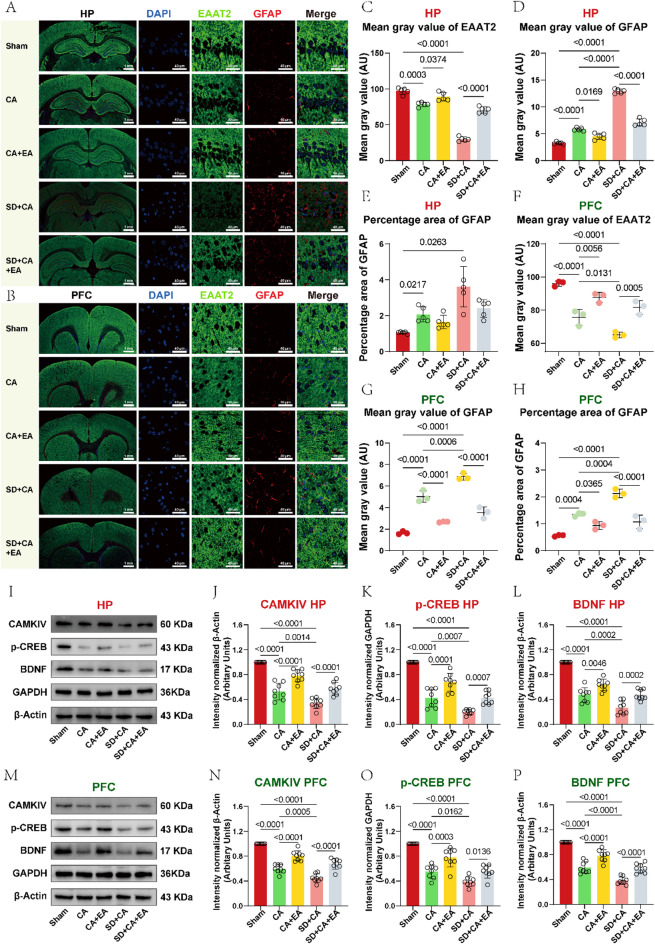
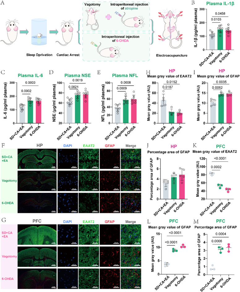
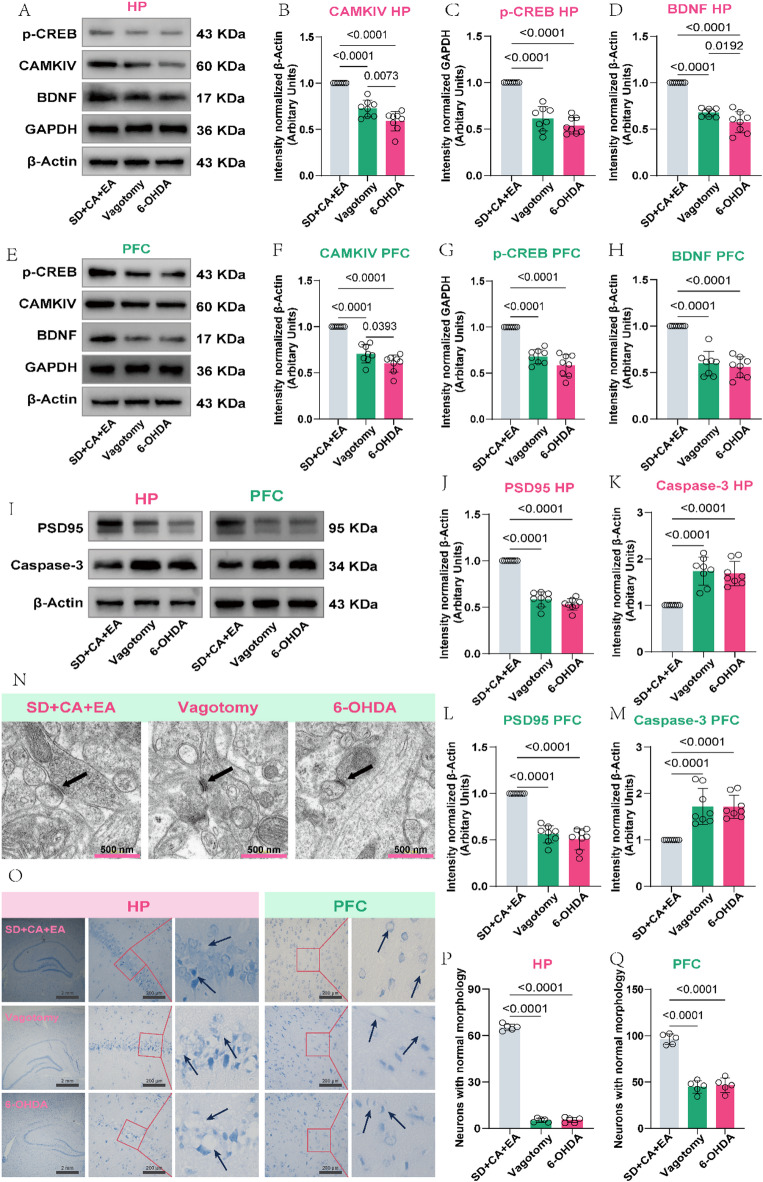
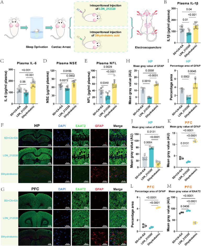
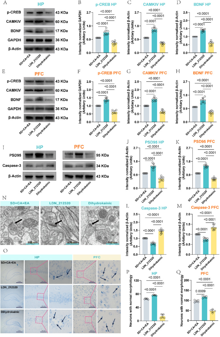
Cardiac arrest (CA)-induced post-cardiac arrest brain injury (PCABI) represents a critical contributor to global mortality and neurological disability. While sleep deprivation (SD) is recognized to aggravate neurological outcomes, its role in PCABI pathogenesis remains underexplored. This study investigated the mechanisms by which SD exacerbates PCABI and evaluated the neuroprotective efficacy of electroacupuncture (EA). A CA model was established in SD rats, followed by RNA sequencing and molecular analyses to assess brain injury biomarkers, synaptic plasticity, and calcium signaling pathways. SD disrupted circadian rhythms, amplified neuronal apoptosis, and suppressed glutamate transporter Excitatory Amino Acid Transporter 2 (EAAT2) expression post-CA, correlating with worsened cognitive deficits. EA treatment significantly attenuated these effects, restoring EAAT2 levels, mitigating calcium overload, and enhancing synaptic integrity. Mechanistically, EA modulated the EAAT2/calcium signaling axis and rebalanced autonomic nervous activity, thereby reducing oxidative stress and neuronal excitotoxicity. These findings identify EAAT2 downregulation as a key mediator of SD-aggravated PCABI and establish EA as a dual-target intervention that rectifies glutamatergic dysregulation and autonomic dysfunction. The study provides translational insights into EA's therapeutic potential for PCABI, particularly in populations with comorbid sleep disturbances.

Keywords: Brain injury; Cardiac arrest; EAAT2; Electroacupuncture; Sleep deprivation.


Conflict of interest statement

Declarations. Competing interests: The authors declare no competing interests. Compliance with ethics requirements: All animal experiment protocols were performed in accordance with the guidelines of Animal Care and Use Committees at the Harbin Medical University (Harbin, Heilongjiang, China) and were in compliance with the Guide for the Care and Use of Laboratory Animals published by the US National Institutes of Health (NIH Publication No. 85 − 23, revised 2011).

Figures

Fig. 1
Fig. 1
Electroacupuncture alleviates post-cardiac arrest brain injury. (A) Flowchart of the experiment. (B-E) Plot of Elisa assays for IL-1β, IL-6, NSE and NFL in the plasma of rats on day 4 post-CA (n = 8). (F) Representative micrographs showing Nissl histochemical staining of hippocampus and prefrontal cortex neurons, alongside TEM of the CA1 hippocampal subfield at 4 days post-CA, black arrows indicate chromatolytic neurons with Nissl body disintegration, while green arrows indicate synaptic regions. (G-H) Nissl body number in hippocampus and prefrontal cortex on day 4 post-CA (n = 5). (I-N) Relative expression of PSD95, and Caspase-3 in hippocampus and prefrontal cortex on day 4 post-CA (n = 8).
Fig. 2
Fig. 2
Electroacupuncture corrects sleep deprivation-induced transcriptomic alterations in post-cardiac arrest hippocampus. (A-C) Volcano plots of differentially expressed RNAs (|log2FC| > 0.58, adjusted p < 0.05) between: (A) CA vs. SD + CA (309 upregulated, 274 downregulated); (B) CA vs. CA + EA (185 upregulated, 127 downregulated); (C) SD + CA vs. SD + CA + EA (220 upregulated, 553 downregulated). Dashed lines indicate significance thresholds. (D-F) KEGG pathway enrichment analysis of DEGs for: (D) CA vs. CA + EA; (E) SD + CA vs. CA; (F) SD + CA vs. SD + CA + EA. Bubble size represents gene count; color indicates -log10(q-value). (G) Protein-protein interaction network of consolidated DEGs (combined SD + CA vs. CA and SD + CA vs. SD + CA + EA comparisons). Hub genes identified by betweenness centrality: Slc1a2, Grin2a, Nos1.n = 8 biologically independent samples per group.
Fig. 3
Fig. 3
Electroacupuncture restores SD-compromised EAAT2/calcium signaling axis in post-cardiac arrest neurodegeneration. (A-B) Representative immunofluorescent images of EAAT2 (green) and astroglial marker GFAP (red) in hippocampal CA1 and prefrontal cortex regions, Nuclei counterstained with DAPI (blue). (C-H) Quantitative analysis of EAAT2 and GFAP fluorescence intensity, and GFAP-positive area fraction in hippocampus and prefrontal cortex. (I, M) Western blot membranes showing calcium signaling proteins (CAMKIV, p-CREB, BDNF) with loading controls (GAPDH/β-actin) in hippocampus and prefrontal cortex lysates. (J-L, N-P) Densitometric quantification of CAMKIV, p-CREB, and BDNF expression levels in hippocampus and prefrontal cortex.
Fig. 4
Fig. 4
Autonomic imbalance abrogates electroacupuncture-mediated EAAT2 restoration in post-cardiac arrest neurodegeneration (A) Experimental timeline for autonomic nerve inhibition (vagotomy/atropine vs. 6-OHDA) combined with EA intervention. (B-E) Plasma biomarker profiles showing elevated IL-1β, IL-6, NSE, and NFL under autonomic dysfunction, despite EA treatment (n = 8). (F-G) Representative confocal images of EAAT2 (green) and astroglial GFAP (red) in hippocampal CA1 and prefrontal cortex regions, nuclei stained with DAPI (blue). (H-J) Quantitative analysis of hippocampal EAAT2 and GFAP fluorescence intensity, and GFAP area fraction (n = 5). (K-M) Quantitative analysis of Prefrontal cortical EAAT2 and GFAP fluorescence intensity, and GFAP area fraction (n = 3).
Fig. 5
Fig. 5
Electroacupuncture fails to ameliorate severe nerve damage in the presence of autonomic imbalance. (A) WB bands of CAMKIV, p-CREB, BDNF, GAPDH and β-Actin in hippocampus. (B-D) Relative expression of CAMKIV, p-CREB, and BDNF in hippocampus (n = 8). (E) WB bands of CAMKIV, p-CREB, BDNF, GAPDH and β-Actin in prefrontal cortex. (F-H) Relative expression of PSD95, CAMKIV, p-CREB, BDNF and Caspase-3 in prefrontal cortex (n = 8). (I) WB bands of PSD95, Caspase-3 and β-Actin in hippocampus and prefrontal cortex. (J-M) Relative expression of PSD95, and Caspase-3 in hippocampus and prefrontal cortex (n = 8). (N) TEM of the CA1 region of the hippocampus, black arrows indicate synaptic regions. (O) Nissl histochemical stain for Nissl bodies in the neurons of hippocampus and prefrontal cortex in different rat groups, black arrows indicate chromatolytic neurons with Nissl body disintegration. (P-Q) Nissl body number in hippocampus and prefrontal cortex (n = 5).
Fig. 6
Fig. 6
Under conditions of autonomic nerve imbalance, the regulatory effect of electroacupuncture on EAAT2 is ineffective. (A) The experimental flow chart. (B-E) Plot of Elisa assays for IL-1β, IL-6, NSE and NFL in the plasma of rats on day 4 post-CA (n = 8). (F-G) EAAT2 and GFAP immunofluorescence staining in hippocampus and prefrontal cortex. (H-J) Immunofluorescence semi-quantitative analysis of EAAT2 and GFAP in hippocampus (n = 5). (K-M) Immunofluorescence analysis of EAAT2 and GFAP in prefrontal cortex (n = 3).
Fig. 7
Fig. 7
Under conditions of autonomic nerve imbalance, electroacupuncture cannot improve PCABI. (A) WB bands of CAMKIV, p-CREB, BDNF, GAPDH and β-Actin in hippocampus. (B-D) Relative expression of CAMKIV, p-CREB, and BDNF in hippocampus (n = 8). (E) WB bands of CAMKIV, p-CREB, BDNF, GAPDH and β-Actin in prefrontal cortex. (F-H) Relative expression of PSD95, CAMKIV, p-CREB, BDNF and Caspase-3 in prefrontal cortex (n = 8). (I) WB bands of PSD95, Caspase-3 and β-Actin in hippocampus and prefrontal cortex. (J-M) Relative expression of PSD95, and Caspase-3 in hippocampus and prefrontal cortex (n = 8). (N) TEM of the CA1 region of the hippocampus, black arrows indicate synaptic regions. (O) Nissl histochemical stain for Nissl bodies in the neurons of hippocampus and prefrontal cortex in different rat groups, black arrows indicate chromatolytic neurons with Nissl body disintegration. (P-Q) Nissl body number in hippocampus and prefrontal cortex (n = 5).

Similar articles

References

    1. Ramireddy, A. S. S. & Chugh Do peak times exist for sudden cardiac arrest? Trends Cardiovasc. Med.31 (3), 172–176 (2021). - PMC - PubMed
    1. Perkins, G. D. et al. Brain injury after cardiac arrest. Lancet398 (10307), 1269–1278 (2021). - PubMed
    1. Sekhon, M. S., Ainslie, P. N. & Griesdale, D. E. Clinical pathophysiology of hypoxic ischemic brain injury after cardiac arrest: A two-hit model. Crit. Care. 21 (1). 10.1186/s13054-017-1670-9 (2017). - PMC - PubMed
    1. Sandroni, C., Cronberg, T. & Sekhon, M. Brain injury after cardiac arrest: pathophysiology, treatment, and prognosis. Intensive Care Med.47 (12), 1393–1414 (2021). - PMC - PubMed
    1. Chen, C. et al. Rna-seq analysis of the key long noncoding Rnas and Mrnas related to cognitive impairment after cardiac arrest and cardiopulmonary resuscitation. Aging (Albany NY). 12 (14), 14490–14505 (2020). - PMC - PubMed

Substances

LinkOut - more resources