. 2025 Aug;29(15):e70772.
doi: 10.1111/jcmm.70772.

Tianma Granules Alleviate AOM/DSS-Induced Colorectal Tumorigenesis by Inhibiting the Wnt/β-Catenin Pathway Activation

Affiliations

Tianma Granules Alleviate AOM/DSS-Induced Colorectal Tumorigenesis by Inhibiting the Wnt/β-Catenin Pathway Activation

Renjing Lin et al. J Cell Mol Med. 2025 Aug.

Abstract

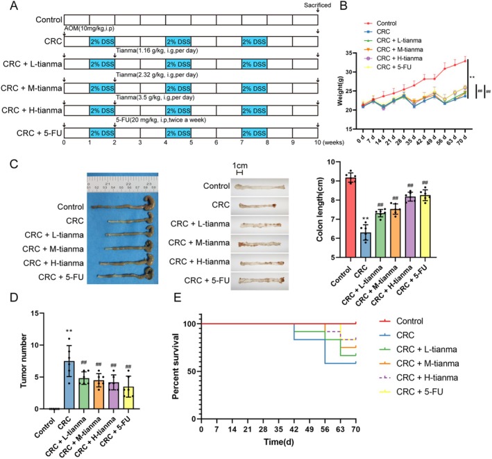
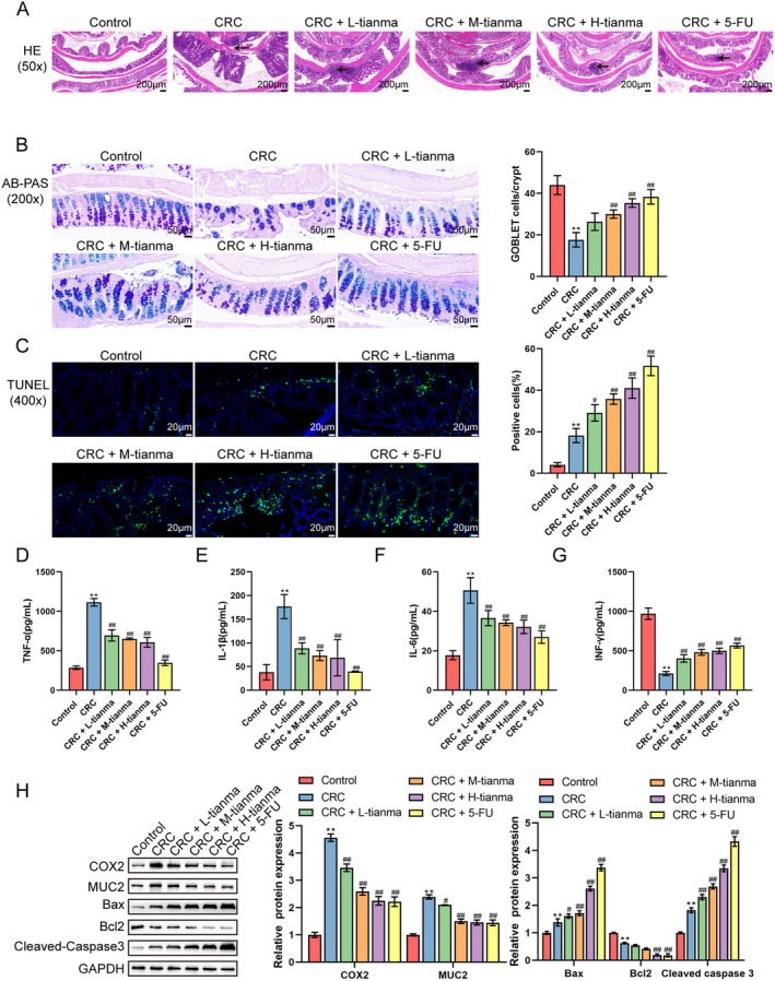
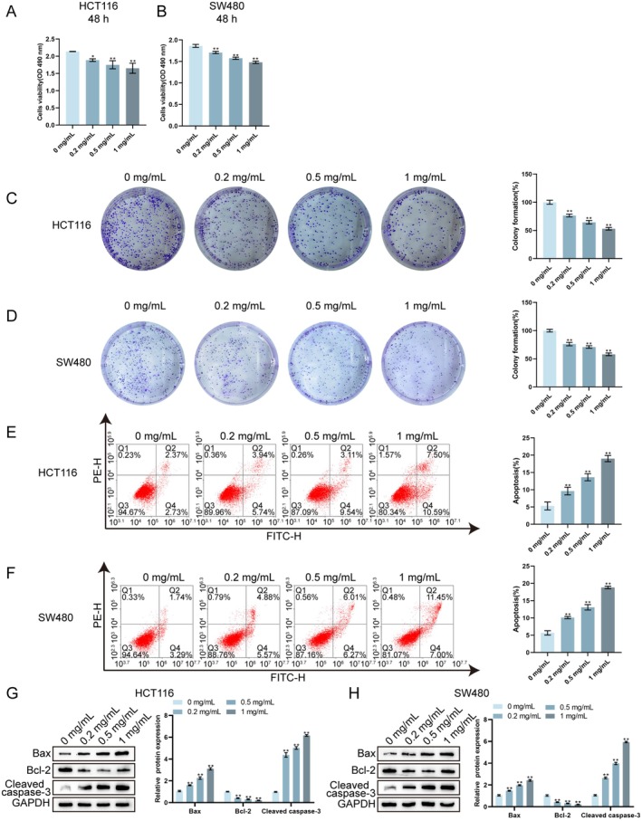
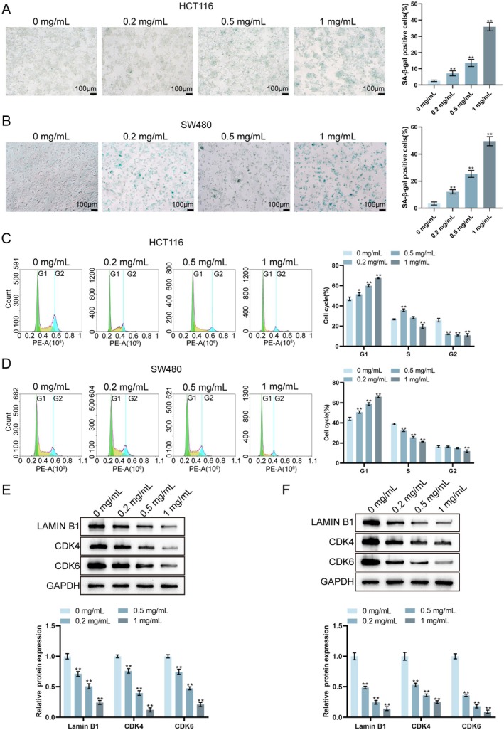
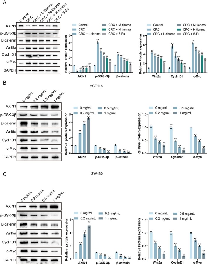
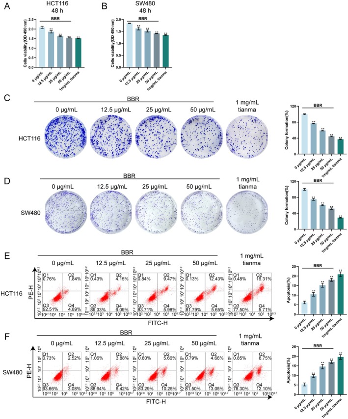
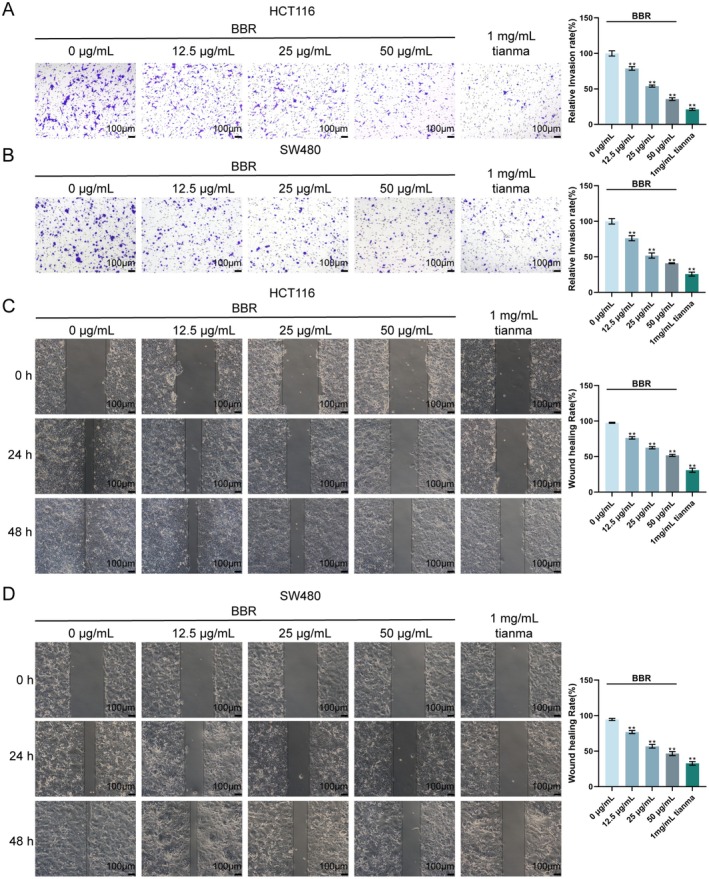
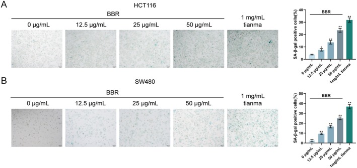
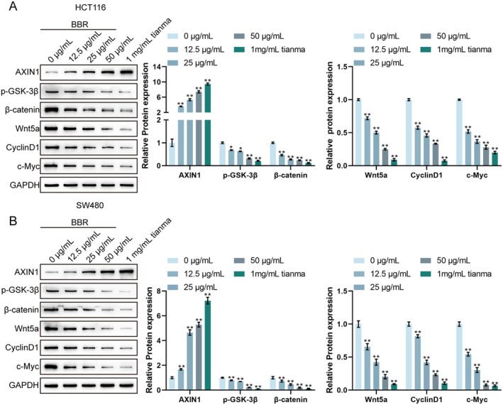
This study aimed to investigate the anti-tumour effect and the possible molecular mechanism of Tianma granules on colorectal cancer (CRC). The therapeutic effect of Tianma granules on CRC cell lines (HT116 and SW480) and AOM/DSS-induced CRC mouse models was evaluated. Tianma granules can attenuate weight loss and increase the survival rate of CRC mice, restore reduced colon length, reduce tumour numbers and increase goblet cell numbers in CRC mice. Tianma granules also downregulated the level of CRC-specific markers (COX2 and MUC2), inhibited the inflammation (decreased TNF-α, IL-1β, IL-6 levels and increased INF-γ level), and promoted apoptosis (decreased TUNEL positive cell rate; decreased Bax and Cleaved caspase3 protein levels and increased Bcl2 level) in CRC mice. In vitro, Tianma granules can inhibit the viability, proliferation, migration and invasion of CRC cells, while promoting cell apoptosis, cell cycle arrest and cell senescence. Tianma granules promoted AXIN1 protein levels and inhibited p-GSK-3β, β-catenin, Wnt5a and Cyclin D1 and c-Myc protein levels. Moreover, the network pharmacology analysis and in vitro validation revealed berberine might be the key compound responsible for Tianma granules' pharmacological actions. In conclusion, Tianma granules can inhibit inflammation and tumour progression in AOM/DSS-induced CRC mice, as well as inhibit CRC cell malignant phenotype. The protection of Tianma granules against CRC may be achieved by inhibiting the Wnt signalling pathway.

Keywords: AOM/DSS; Tianma granules; Wnt signalling pathway; colorectal cancer; inflammation.


Conflict of interest statement

The authors declare no conflicts of interest.

Figures

FIGURE 1
FIGURE 1
Tianma granules can improve the occurrence and development of CRC in AOM/DSS‐induced mice. (A) Schematic diagram of AOM/DSS model experimental program; (B) changes in mouse body weight were analysed; (C) colon length and (D) CRC tumour numbers in mice were analysed; (E) mouse survival rate was detected. N = 6. Data are expressed as mean ± SD. Statistical significance was determined by one‐way ANOVA followed by Tukey's multiple comparisons test for (B–D). Survival rate in (E) was analysed by Kaplan–Meier survival curves and log‐rank test. **p < 0.01, compared with the control group; ## p < 0.01, compared with the CRC group.
FIGURE 2
FIGURE 2
Tianma granules alleviated the pathological damage and inflammation of CRC mice and promoted CRC cell apoptosis. (A) H&E staining was used to detect the pathological damage of colon tissues (black arrows indicated tumours in the colon). (B) AB‐PAS staining was used to detect and quantify goblet cell numbers in colon tissues. (C) TUNEL staining was used to detect cell apoptosis and the apoptotic cell rate in colon tumour tissues (green fluorescence represented apoptotic cells). (D–G) ELISA was used to detect the levels of inflammatory cytokines (TNF‐α, IL‐1β, IL‐6 and IFN‐γ) in colon tissues. (H) Western blot was used to detect the protein levels of CRC‐specific proteins (COX2 and MUC2) and apoptotic proteins (Bax, Cleaved‐caspase3 and Bcl2). N = 3. Data are expressed as mean ± SD. Statistical significance was determined by one‐way ANOVA followed by Tukey's multiple comparisons test. **p < 0.01, compared with the control group; # p < 0.05, ## p < 0.01, compared with the CRC group.
FIGURE 3
FIGURE 3
Tianma granules can inhibit CRC cell proliferation and promote cell apoptosis in vitro. After treating HCT116 and SW480 cells with different concentrations of Tianma granules (0.2, 0.5 and 1 mg/mL) for 48 h, MTT assay was used to detect the cell viability of HCT116 cells (A, B) and SW480 cells; (C, D) colony formation assay was used to detect CRC cell proliferation; (E, F) flow cytometry was used to detect CRC cell apoptosis; (G, H) western blot was used to detect the protein levels of Bax, cleaved caspase3 and Bcl‐2 in CRC cells. N = 3. Data are expressed as mean ± SD. Statistical significance was determined by one‐way ANOVA followed by Tukey's multiple comparisons test. *p < 0.05, **p < 0.01.
FIGURE 4
FIGURE 4
Tianma granules inhibited CRC cell migration and invasion in vitro. After treating HCT116 and SW480 cells with different concentrations of Tianma granules (0.2, 0.5 and 1 mg/mL) for 24 h, Transwell assay was used to detect the invasion of (A) HCT116 cells and (B) SW480 cells; scratch assay was used to detect the migration ability of (C) HCT116 cells and (D) SW480 cells. N = 3. Data are expressed as mean ± SD. Statistical significance was determined by one‐way ANOVA followed by Tukey's multiple comparisons test. **p < 0.01.
FIGURE 5
FIGURE 5
Tianma granules promoted CRC cell senescence and cell cycle arrest in vitro. (A, B) HCT116 (A) and SW480 (B) cells were treated with Tianma granules at concentrations of 0, 0.2, 0.5 and 1 mg/mL. Representative images are shown on the left and quantitative analysis of SA‐β‐gal‐positive cells is shown on the right. (C, D) Flow cytometry was used to analyse the cell cycle distribution in HCT116 (C) and SW480 (D) cells treated with different concentrations of Tianma granules. The percentages of cells in the G1, S and G2 phases are shown in the histograms on the right. (E, F) HCT116 (E) and SW480 (F) cells were treated with Tianma granules at the indicated concentrations and the protein levels of lamin B1, CDK4 and CDK6 were determined by western blot. N = 3. Data are expressed as mean ± SD. Statistical significance was determined by one‐way ANOVA followed by Tukey's multiple comparisons test. *p < 0.05, **p < 0.01.
FIGURE 6
FIGURE 6
Tianma granules inhibited the activation of the Wnt pathway in CRC mice and cells. (A) Western blot analysis was used to detect the protein levels of AXIN1, p‐GSK‐3β, β‐catenin, Wnt5a, CyclinD1 and c‐Myc in the colon tissues of AOM/DSS‐induced CRC mice. (B) After treating HCT116 cells with different concentrations of Tianma granules (0.2, 0.5 and 1 mg/mL) for 24 h, western blot analysis was used to detect the protein levels of AXIN1, p‐GSK‐3β, β‐catenin, Wnt5a, CyclinD1 and c‐Myc. (C) Western blot analysis was performed to detect the protein levels of AXIN1, p‐GSK‐3β, β‐catenin, Wnt5a, CyclinD1 and c‐Myc in SW480 cells treated with different concentrations of Tianma granules (0.2, 0.5 and 1 mg/mL) for 24 h. GAPDH served as a loading control. N = 3. Data are expressed as mean ± SD. Statistical significance was determined by one‐way ANOVA followed by Tukey's multiple comparisons test. **p < 0.01, compared with the control/0 mg/mL group; ## p < 0.01, compared with the CRC group.
FIGURE 7
FIGURE 7
Network pharmacology analysis of Tianma granules for colorectal cancer treatment. (A) Volcano plot of differentially expressed genes in the GSE10950 dataset, showing upregulated (up) and downregulated (down) genes with ∣logFC∣ > 1 and p < 0.05. (B) Heatmap illustrating the expression profile of differentially expressed genes in GSE10950 colorectal cancer (tumor) and paired normal colon tissue (normal) samples. (C) Venn diagram showing the overlap among differentially expressed genes from GSE10950, predicted drug‐target genes from Tianma granules (TMKLJ), and colorectal cancer‐related genes. (D) KEGG pathway enrichment analysis of the common target genes. (E) Gene Ontology (GO) enrichment analysis of the common target genes. (F) Protein–protein interaction (PPI) network of the common target genes, with node size representing the degree of connectivity. (G) Expression levels of hub genes (TP53, MYC, CTNNB1) in colorectal cancer (tumour) and normal (normal) samples from the GSE10950 dataset. (H) Sankey diagram illustrating the relationship between key active components of Tianma granules, their hub targets, and relevant signalling pathways in colorectal cancer. The width of the connecting lines corresponds to the strength of the association.
FIGURE 8
FIGURE 8
Berberine inhibits CRC cell proliferation and promotes apoptosis in vitro HCT116 and SW480 cells were treated with different concentrations of berberine (BBR: 12.5, 25 and 50 μg/mL) or 1 mg/mL Tianma granules. (A, B) Cell viability of HCT116 and SW480 cells after 48 h treatment, as determined by MTT assay. (C, D) Colony formation assay results for HCT116 and SW480 cells, showing representative images (left) and quantification of colony formation rate (right). (E) Flow cytometry analysis of apoptosis in HCT116 and SW480 cells, showing representative plots (left) and quantification of apoptotic percentage (right). N = 3. Data are expressed as mean ± SD. Statistical significance was determined by one‐way ANOVA followed by Tukey's multiple comparisons test. **p < 0.01, compared with the 0 μg/mL BBR group.
FIGURE 9
FIGURE 9
Berberine inhibits CRC cell migration and invasion in vitro. (A, B) Transwell assay results for HCT116 and SW480 cell invasion, showing representative images (left) and quantification of relative invasion rate (right). (C, D) Scratch assay results for HCT116 cell and SW480 cell migration, showing representative images at 0, 24 and 48 h (left) and quantification of wound healing rate at 48 h (right). N = 3. Data are expressed as mean ± SD. Statistical significance was determined by one‐way ANOVA followed by Tukey's multiple comparisons test. **p < 0.01, compared with the 0 μg/mL BBR group.
FIGURE 10
FIGURE 10
Berberine promotes CRC cell senescence in vitro. (A, B) Senescence‐associated β‐galactosidase (SA‐β‐gal) staining in HCT116 and SW480 cells, showing representative images (left) and quantification of SA‐β‐gal positive cells (right). N = 3. Data are expressed as mean ± SD. Statistical significance was determined by one‐way ANOVA followed by Tukey's multiple comparisons test. * p < 0.05, **p < 0.01, compared with the 0 μg/mL BBR group.
FIGURE 11
FIGURE 11
Berberine inhibits the activation of the Wnt/β‐catenin pathway in CRC cells. (A) Western blot analysis of Wnt/β‐catenin pathway‐related proteins (AXIN1, p‐GSK‐3β, β‐catenin, Wnt5a, CyclinD1 and c‐Myc) in HCT116 cells and SW480 cells, showing representative blots (left) and quantification of relative protein expression (right). GAPDH served as a loading control. N = 3. Data are expressed as mean ± SD. Statistical significance was determined by one‐way ANOVA followed by Turkey's multiple comparisons test. * p < 0.05, **p < 0.01, compared with the 0 μg/mL BBR group.

Similar articles

References

    1. Li J., Yuan Y., Yang F., et al., “Expert Consensus on Multidisciplinary Therapy of Colorectal Cancer With Lung Metastases (2019 Edition),” Journal of Hematology & Oncology 12, no. 1 (2019): 16. - PMC - PubMed
    1. Marmol I., Sánchez‐de‐Diego C., Pradilla Dieste A., Cerrada E., and Rodriguez Yoldi M. J., “Colorectal Carcinoma: A General Overview and Future Perspectives in Colorectal Cancer,” International Journal of Molecular Sciences 18, no. 1 (2017): 197. - PMC - PubMed
    1. Zhang L., Cao F., Zhang G., et al., “Trends in and Predictions of Colorectal Cancer Incidence and Mortality in China From 1990 to 2025,” Frontiers in Oncology 9 (2019): 98. - PMC - PubMed
    1. Sandhu J., Lavingia V., and Fakih M., “Systemic Treatment for Metastatic Colorectal Cancer in the Era of Precision Medicine,” Journal of Surgical Oncology 119, no. 5 (2019): 564–582. - PubMed
    1. Sethy C. and Kundu C. N., “5‐Fluorouracil (5‐FU) Resistance and the New Strategy to Enhance the Sensitivity Against Cancer: Implication of DNA Repair Inhibition,” Biomedicine & Pharmacotherapy 137 (2021): 111285. - PubMed

MeSH terms