. 2025 Aug;29(15):e70775.
doi: 10.1111/jcmm.70775.

FoxO3 Activation Alleviates Doxorubicin-Induced Cardiomyopathy by Enhancing Autophagic Flux and Suppressing mTOR/ROS Signalling

Affiliations

FoxO3 Activation Alleviates Doxorubicin-Induced Cardiomyopathy by Enhancing Autophagic Flux and Suppressing mTOR/ROS Signalling

Zao-Shang Chang et al. J Cell Mol Med. 2025 Aug.

Abstract

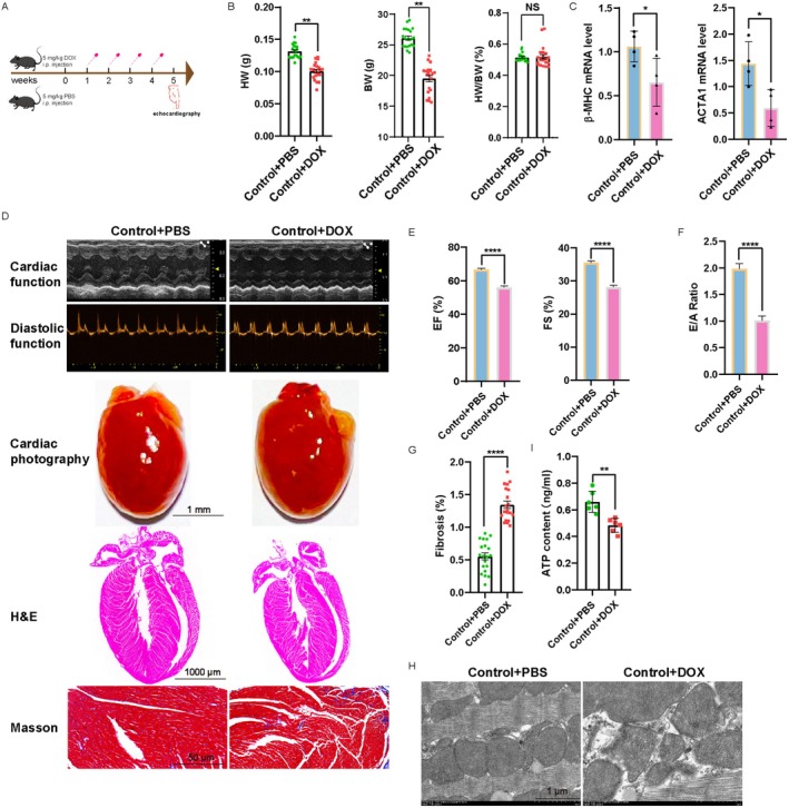
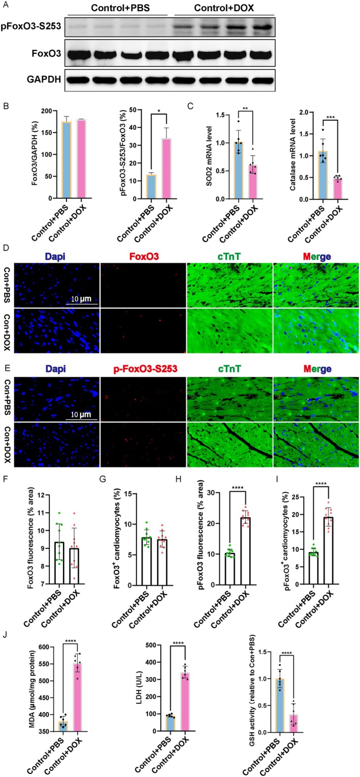
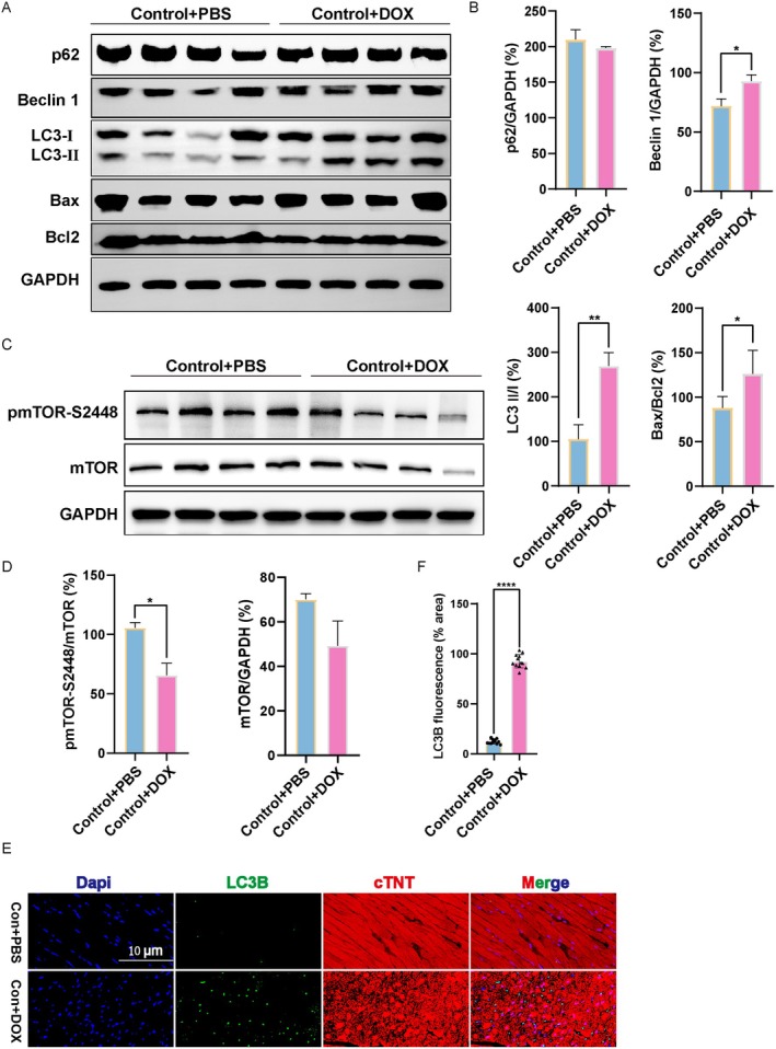
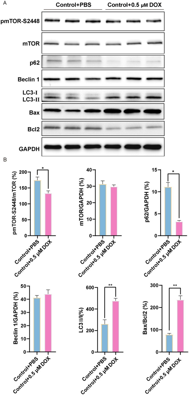
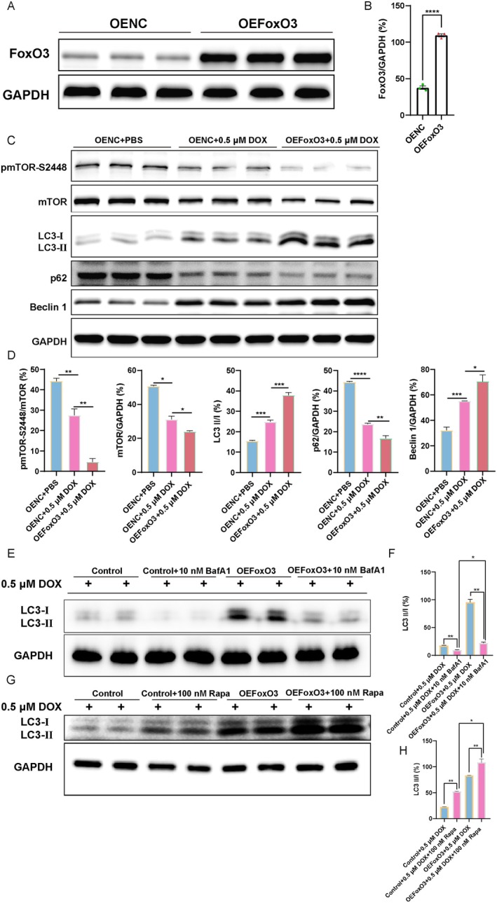
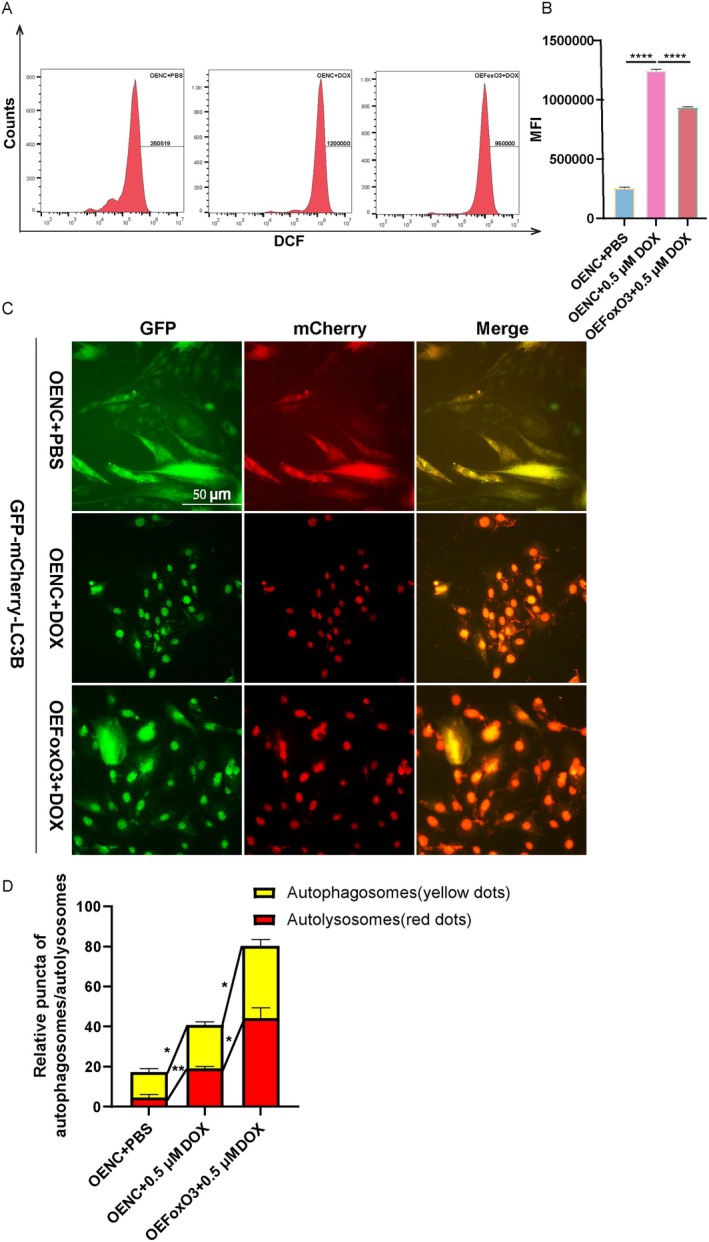
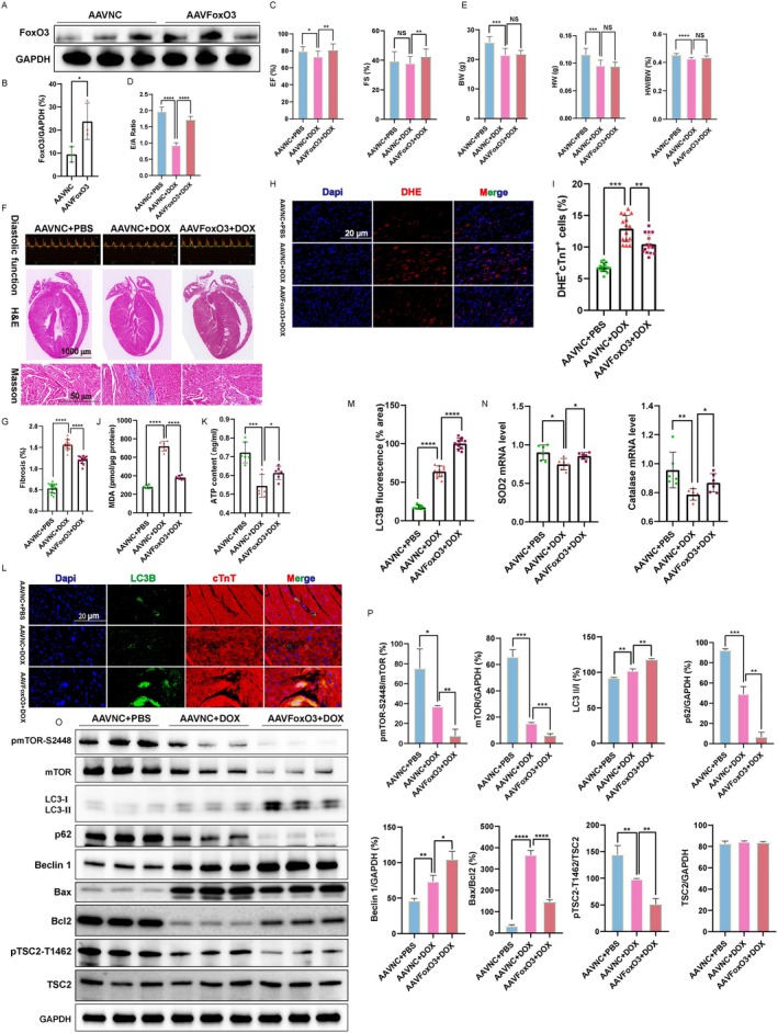
Doxorubicin (DOX) is an effective chemotherapy drug, but its use is limited by cardiotoxicity, known as DOX-induced cardiomyopathy. The transcription factor FoxO3, which regulates autophagy and oxidative stress, has unclear mechanisms in this condition. We found that DOX-induced cardiomyopathy involved cardiac atrophy, cardiac dysfunction, fibrosis and mitochondrial damage. DOX reduced H9c2 cardiomyocyte viability and glutathione levels (GSH), increased reactive oxygen species (ROS), malondialdehyde (MDA) and lactate dehydrogenase (LDH) and inhibited superoxide dismutase 2 (SOD2) and catalase (CAT) expression. DOX also suppressed FoxO3 activation and increased the autophagy protein LC3 II/I ratio. Overexpressing FoxO3 enhanced LC3B, Beclin 1 and autophagic flux, while reducing p62 and suppressing mTOR activation in heart. Brefeldin A1 (BafA1), an autophagy inhibitor and rapamycin (Rapa), an autophagy activator, were administered to H9c2 cardiomyocytes to elucidate the regulatory mechanism of FoxO3. Mechanically, our data revealed that FoxO3 overexpression enhanced autophagy and suppressed ROS production and mTOR activation in both in vitro and in vivo models of DOX exposure. Collectively, targeting FoxO3 to enhance protective autophagy may offer a therapeutic strategy against DOX-induced cardiomyopathy.

Keywords: FoxO3; autophagy; cardiomyopathy; doxorubicin; reactive oxygen species.


Conflict of interest statement

The authors declare no conflicts of interest.

Figures

FIGURE 1
FIGURE 1
DOX induces cardiac atrophy and cardiac dysfunction in vivo. (A) Schematic diagram depicting the protocol for DOX and PBS intraperitoneal injection; PBS was used as negative control. The figure was drawn by Figdraw. (B) Changes in heart weight (HW), body weight (BW) and the ratio of HW/BW (n = 21 for each group). (C) β‐MHC and ACTA1 were analysed by qRT‐PCR (n = 4 hearts for each group). (D) Cardiac function was evaluated by echocardiography (top panel). Representative images of cardiac photography, H&E staining and Masson staining. (E) The ejection fractions (EF) and fraction shortening (FS) were calculated by M‐mode echocardiography (n = 15 for each group). (F) The E/A ratio was calculated (n = 15 for each group). (G) Cardiac fibrosis was quantified after Masson trichrome staining (n = 20 hearts per group). (H) Transmission electron microscopy (TEM) images of heart tissues from each group (n = 3 for each group). (I) ATP content in myocardial tissue was quantified (n = 6 for each group). Data are presented as mean ± SEM, *p < 0.05, **p < 0.01 and ****p < 0.0001.
FIGURE 2
FIGURE 2
FoxO3 transcriptional activity and the expression of antioxidant genes are suppressed in DOX cardiomyopathy. (A) Representative images of western blot for FoxO3 and phospho‐FoxO3‐Ser253. (B) FoxO3 expression and phospho‐FoxO3/total FoxO3 ratio (n = 4). (C) SOD2 and CAT mRNA expression (n = 6). (D‐E) FoxO3 and p‐FoxO3‐S253 expression were detected by immunofluorescence staining. (F‐G) Quantification of FoxO3 fluorescence intensity and the FoxO3+ cTnT+ cells (n = 10 per group). (H‐I) Quantification of p‐FoxO3‐S253 fluorescence intensity and the p‐FoxO3‐S253+ cTnT+ cells (n = 10 per group). (J) MDA, LDH and GSH enzyme activity in cardiac tissue were quantified (n = 6 per group). Data are presented as Mean ± SEM, *p < 0.05, **p < 0.01, ***p < 0.001 and ****p < 0.0001.
FIGURE 3
FIGURE 3
Effects of DOX on autophagy and mTOR in heart tissue. (A) Western blot images of p62, Beclin 1, LC3 II/I, Bax and Bcl2. (B) The relative expression levels of target proteins were quantified as the percentage of Control group (n = 4). (C) Western blot images of mTOR and p‐mTOR‐Ser2448. (D) The relative expression levels of target proteins were quantified as the percentage of Control (n = 4). (E) The localisation and expression of LC3B in myocardium were detected by immunofluorescence staining. Representative images of LC3B were shown (green). cTnT, cardiomyocytes marker. (F) Quantification of LC3B fluorescence intensity was performed (n = 12 per group). Data are presented as Mean ± SEM, *p < 0.05, **p < 0.01 and ****p < 0.0001.
FIGURE 4
FIGURE 4
Effects of DOX on autophagy, mTOR and apoptosis in H9c2 cardiomyocytes. (A) Western blot images of p‐mTOR‐Ser2448, mTOR, p62, Beclin 1, LC3 II/I, Bax and Bcl2 in H9c2 cardiomyocytes. (B) The relative expression levels of target proteins were quantified as the percentage of the Control group (n = 3). Data are presented as Mean ± SEM, *p < 0.05 and **p < 0.01.
FIGURE 5
FIGURE 5
Overexpression of FoxO3 increases autophagy and inhibits activation of mTOR in H9c2 cardiomyocytes. (A‐B) Validation of FoxO3 overexpression (n = 3). (C) Western blot images of p‐mTOR‐Ser2448, mTOR, p62, Beclin 1 and LC3 II/I. (D) The relative expression levels of target proteins were quantified as the percentage of Control (n = 3). (E) Western blot images of LC3 II/I. (F) The relative expression levels of target proteins were quantified as the percentage of Control group. (G) Western blot images of LC3 II/I. (H) The relative expression levels of target proteins were quantified as the percentage of Control group. Data are presented as Mean ± SEM, *p < 0.05, **p < 0.01, ***p < 0.001 and ****p < 0.0001.
FIGURE 6
FIGURE 6
Overexpression of FoxO3 inhibits intracellular ROS and enhances autophagy flow. (A) ROS detection by CM‐H2DCFDA in flow cytometry. (B) Quantification of mean fluorescence intensity (MFI) (n = 3). (C‐D) mCherry‐red fluorescent protein‐green fluorescent protein (GFP)‐LC3B adenovirus was transfected into H9c2 cardiomyocytes to observe changes in autophagic flux. The yellow puncta and red puncta represent autophagosomes and autolysosomes, respectively, which were counted on 30 cells (n = 3 for each group). Data are presented as Mean ± SEM, *p < 0.05, **p < 0.01 and ****p < 0.0001.
FIGURE 7
FIGURE 7
Overexpression of FoxO3 protects DOX‐induced cardiomyopathy by activating autophagy. (A‐B) Validation of FoxO3 overexpression in hearts (n = 3). (C) The ejection fractions (EF) and fraction shortening (FS) were calculated by M‐mode echocardiography (n = 20 for each group). (D) Diastolic function (E/A ratio) was calculated (n = 20 for each group). (E) Changes of heart weight (HW), body weight (BW) and the ratio of HW/BW in the AAVNC+PBS, AAVNC+DOX and AAVFoxO3 + DOX groups (n = 15 for each group). (F) At the end of the experiment, diastolic function was tested (top panel). Representative images of H&E staining and Masson staining. (G) Quantitative analysis of myocardial fibrosis (n = 15 for per group). (H) Representative images of dihydroethidium (DHE) staining (red). (I) Quantification of DHE+ cTnT+ cells in hearts (n = 15 for each group). (J) Quantification of MDA concentration in hearts (n = 6 hearts for each group). (K) Quantification of ATP productions in hearts (n = 6 hearts for each group). (L) The localisation and expression of LC3B in myocardium were detected by immunofluorescence staining. Representative images of LC3B were shown (green). cTnT, cardiomyocytes marker (n = 10 for each group). (M) Quantification of LC3B fluorescence intensity was performed. (N) Cardiac SOD2 and CAT mRNA expression (n = 6). (O) Western blot images of p‐mTOR‐Ser2448, mTOR, p62, Beclin 1, LC3 II/I, Bax, Bcl2, p‐TSC2‐Thr1462 and TSC2. (P) The relative expression levels of target proteins were quantified (n = 3). Data are presented as Mean ± SEM, *p < 0.05, **p < 0.01, ***p < 0.001 and ****p < 0.0001.

Similar articles

References

    1. Zamorano J. L., Lancellotti P., Rodriguez Munoz D., et al., “2016 ESC Position Paper on Cancer Treatments and Cardiovascular Toxicity Developed Under the Auspices of the ESC Committee for Practice Guidelines: The Task Force for Cancer Treatments and Cardiovascular Toxicity of the European Society of Cardiology (ESC),” European Journal of Heart Failure 19, no. 1 (2017): 9–42, 10.1002/ejhf.654. - DOI - PubMed
    1. Lopez‐Sendon J., Alvarez‐Ortega C., Zamora Aunon P., et al., “Classification, Prevalence, and Outcomes of Anticancer Therapy‐Induced Cardiotoxicity: The CARDIOTOX Registry,” European Heart Journal 41, no. 18 (2020): 1720–1729, 10.1093/eurheartj/ehaa006. - DOI - PubMed
    1. Kobza C., “Cardiac Toxicity: Using Angiotensin‐Converting Enzyme Inhibitors to Prevent Anthracycline‐Induced Left Ventricular Dysfunction and Cardiomyopathy,” Clinical Journal of Oncology Nursing 25, no. 3 (2021): 259–266, 10.1188/21.CJON.259-266. - DOI - PubMed
    1. Shakir D. K. and Rasul K. I., “Chemotherapy Induced Cardiomyopathy: Pathogenesis, Monitoring and Management,” Journal of Clinical Medical Research 1, no. 1 (2009): 8–12, 10.4021/jocmr2009.02.1225. - DOI - PMC - PubMed
    1. Huang C., Zhang X., Wang S., et al., “PARP‐2 Mediates Cardiomyocyte Aging and Damage Induced by Doxorubicin Through SIRT1 Inhibition,” Apoptosis 29, no. 5–6 (2024): 816–834, 10.1007/s10495-023-01929-y. - DOI - PubMed

MeSH terms