Arm Position with Increased Risk of Partial Subscapularis Tear Progression Owing to Subluxation of the Long Head of Biceps Tendon: Cadaveric Biomechanical Study
- PMID: 40785757
- PMCID: PMC12328111
- DOI: 10.4055/cios24233
Arm Position with Increased Risk of Partial Subscapularis Tear Progression Owing to Subluxation of the Long Head of Biceps Tendon: Cadaveric Biomechanical Study
Abstract
Backgroud: This study aimed to evaluate the differences in long head of biceps (LHB) motion between the normal and subscapularis intrasubstance partial tear conditions and identify the arm positions that exhibit the most significant LHB motion differences using a cadaveric biomechanical study.
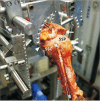
Methods: The LHB tendons of 6 fresh-frozen cadaveric shoulders (2 men and 4 women; mean age, 68.4 ± 2.3 years; range, 65-71 years) were marked with metal beads and mounted in a custom-made shoulder testing system. Data for arm positions at 20° or 60° of forward flexion or abduction, with neutral rotation and maximum internal and external rotation with a torque of 1.5 N·m, were collected. Considering the scapulohumeral rhythm, 20° or 60° forward flexion or abduction in a cadaveric shoulder corresponds to 30° or 90° shoulder elevation in vivo . Mediolateral (subluxation) and inferosuperior (excursion) LHB motions were measured using a 3-dimensional digitizer, and the differences between normal and subscapularis partial tear conditions were analyzed.
Results: While the LHB mediolateral motion difference was the highest during 60° forward flexion with neutral rotation (1.2 ± 0.4, p = 0.042), 20° forward flexion with neutral rotation (0.9 ± 0.3, p = 0.024) and 60° abduction with maximum external rotation (0.9 ± 0.3, p = 0.036) also demonstrated high mediolateral LHB motion difference between the normal and subscapularis partial tear conditions. In contrast, the LHB inferosuperior motion difference was the highest during 20° forward flexion with neutral rotation (0.7 ± 0.3, p = 0.045) between the normal and subscapularis partial tear conditions.
Conclusions: Upon comparing normal and subscapularis partial tear conditions in this cadaveric study, high pathological movements of the LHB were observed during arm forward flexion with neutral rotation and abduction with external rotation. Repetitive activity in these arm positions could aggravate the condition in a partial subscapularis tear.
Keywords: Biomechanic; Cadaver; Rotator cuff Injuries; Shoulder Impingement syndrome.
Copyright © 2025 by The Korean Orthopaedic Association.
Conflict of interest statement
CONFLICT OF INTEREST: No potential conflict of interest relevant to this article was reported.
Figures


Similar articles
-
Releasing Forces in Adhesive Capsulitis Are Important Indicators of Shoulder Stiffness and Postoperative Function.Clin Orthop Relat Res. 2025 Jun 1;483(6):1033-1046. doi: 10.1097/CORR.0000000000003365. Epub 2025 Jan 28. Clin Orthop Relat Res. 2025. PMID: 39887151
-
Biomechanical effects of different rotator cuff tear conditions and mediolateral offset in lateralized reverse total shoulder arthroplasty (Coralis Reverse Shoulder System).J Shoulder Elbow Surg. 2025 May 6:S1058-2746(25)00348-9. doi: 10.1016/j.jse.2025.03.027. Online ahead of print. J Shoulder Elbow Surg. 2025. PMID: 40339783
-
Biomechanical evaluation of shear force vectors leading to injury of the biceps reflection pulley: a biplane fluoroscopy study on cadaveric shoulders.Am J Sports Med. 2010 May;38(5):1015-24. doi: 10.1177/0363546509355142. Epub 2010 Mar 22. Am J Sports Med. 2010. PMID: 20308434
-
Arthroscopic Repair of Isolated Subscapularis Tears: A Systematic Review of Technique-Specific Outcomes.Arthroscopy. 2017 Apr;33(4):849-860. doi: 10.1016/j.arthro.2016.10.020. Epub 2017 Jan 9. Arthroscopy. 2017. PMID: 28082063
-
Subscapularis repair techniques for reverse total shoulder arthroplasty: A systematic review.J ISAKOS. 2022 Dec;7(6):181-188. doi: 10.1016/j.jisako.2022.05.001. Epub 2022 May 18. J ISAKOS. 2022. PMID: 35597429
References
-
- Song HE, Jang SH, Kim JG. Comparison of two arthroscopic coracoplasty approaches in subscapularis tears. Clin Should Elbow. 2017;20(4):189–194.
-
- Hackl M, Buess E, Kammerlohr S, et al. A “comma sign”-directed subscapularis repair in anterosuperior rotator cuff tears yields biomechanical advantages in a cadaveric model. Am J Sports Med. 2021;49(12):3212–3217. - PubMed
MeSH terms
LinkOut - more resources
Full Text Sources
Medical

