

Intravoxel incoherent motion diffusion-weighted imaging for the assessment of renal injury in cirrhotic patients
- PMID: 40785862
- PMCID: PMC12332735
- DOI: 10.21037/qims-2024-2918
Intravoxel incoherent motion diffusion-weighted imaging for the assessment of renal injury in cirrhotic patients
Abstract
Background: Renal dysfunction is a common complication in patients with cirrhosis, and early detection is crucial for timely intervention and treatment. Intravoxel incoherent motion (IVIM) diffusion-weighted imaging (DWI) serves as a non-invasive imaging technique that provides valuable insights into tissue perfusion and diffusion changes, demonstrating significant superiority in assessing renal injury. This prospective study aimed to evaluate early renal injury in patients with cirrhosis using IVIM DWI and to explore the correlation of IVIM parameters with the severity of liver cirrhosis based on the Child-Pugh classification.
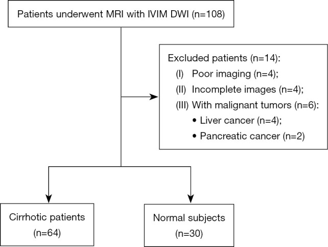
Methods: Sixty-four cirrhotic patients and 30 healthy subjects underwent IVIM on a 3.0-T magnetic resonance imaging (MRI). Diffusion coefficient (ADCslow), pseudo-diffusion coefficient (ADCfast), and perfusion fraction (f) were derived from the bi-exponential model. In the control group, IVIM-derived parameters for both renal cortex and medulla were compared between right and left kidneys. Subsequently, IVIM-derived renal cortical and medullary parameters were compared between the cirrhotic and control groups. Additionally, within the cirrhotic group, IVIM-derived renal parameters were correlated with the Child-Pugh classification.
Results: In the control group, bilateral renal IVIM parameters (ADCslow, ADCfast, and f) showed no significant differences between left and right kidneys (all P>0.05), justifying the use of averaged values for subsequent analysis. Comparative assessment between cirrhotic patients and controls revealed significantly lower renal cortical ADCfast (P=0.033) and f values (P=0.049) in the cirrhotic group, while ADCslow did not differ significantly (P=0.846). In the renal medulla, cirrhotic patients exhibited reduced ADCfast (P=0.043) compared with controls, with no differences in ADCslow or f (P=0.638 and 0.173, respectively). Notably, renal cortical ADCfast demonstrated a significant inverse correlation with Child-Pugh classification (R=-0.406, P=0.001), being higher in Child-Pugh A vs. B (P=0.032) and A vs. C (P=0.019) patients, whereas ADCslow and f showed no correlation with hepatic function grade (P=0.817 and 0.191). Similarly, renal medullary ADCfast correlated inversely with Child-Pugh classification (R=-0.251, P=0.045), with higher values in class A vs. B (P=0.017), but no differences between class A vs. C (P=0.052) or B vs. C (P=0.448). ADCslow and f values in both cortical and medullary regions remained non-significant across Child-Pugh classification (all P>0.05).
Conclusions: IVIM DWI non-invasively assesses early renal injury in cirrhotic patients, with reduced renal perfusion correlating with liver cirrhosis severity.
Keywords: Cirrhosis; diffusion-weighted imaging (DWI); intravoxel incoherent motion (IVIM); kidneys; magnetic resonance imaging (MRI).
Copyright © 2025 AME Publishing Company. All rights reserved.
Conflict of interest statement
Conflicts of Interest: All authors have completed the ICMJE uniform disclosure form (available at https://qims.amegroups.com/article/view/10.21037/qims-2024-2918/coif). R.H. reports that this research was funded by the Scientific and Technological Research Program of Chongqing Municipal Education Commission (No. KJQN202415136). Hua Yang reports that this research was funded by the Natural Science Foundation of Chongqing, China (No. CSTB2024NSCQ-MSX0230). L.N. is an employee of GE Healthcare, MR Research China, Beijing, China. The other authors have no conflicts of interest to declare.
Figures


Similar articles
-
Characterizing Breast Tumor Heterogeneity Through IVIM-DWI Parameters and Signal Decay Analysis.Diagnostics (Basel). 2025 Jun 12;15(12):1499. doi: 10.3390/diagnostics15121499. Diagnostics (Basel). 2025. PMID: 40564820 Free PMC article.
-
Intravoxel incoherent motion diffusion-weighted imaging for evaluating the pancreatic perfusion in cirrhotic patients.Abdom Radiol (NY). 2024 Feb;49(2):492-500. doi: 10.1007/s00261-023-04063-0. Epub 2023 Dec 5. Abdom Radiol (NY). 2024. PMID: 38052890
-
Experimental study of intravoxel incoherent motion diffusion imaging combined with ultrasound renal resistance index in contrast-induced nephropathy.BMC Nephrol. 2025 Jul 18;26(1):401. doi: 10.1186/s12882-025-04329-3. BMC Nephrol. 2025. PMID: 40682007 Free PMC article.
-
Advanced breast diffusion-weighted imaging: what are the next steps? A proposal from the EUSOBI International Breast Diffusion-weighted Imaging working group.Eur Radiol. 2025 Apr;35(4):2130-2140. doi: 10.1007/s00330-024-11010-0. Epub 2024 Oct 8. Eur Radiol. 2025. PMID: 39379708 Free PMC article. Review.
-
Magnetic resonance perfusion for differentiating low-grade from high-grade gliomas at first presentation.Cochrane Database Syst Rev. 2018 Jan 22;1(1):CD011551. doi: 10.1002/14651858.CD011551.pub2. Cochrane Database Syst Rev. 2018. PMID: 29357120 Free PMC article.
References
-
- Tandon P, James MT, Abraldes JG, Karvellas CJ, Ye F, Pannu N. Relevance of New Definitions to Incidence and Prognosis of Acute Kidney Injury in Hospitalized Patients with Cirrhosis: A Retrospective Population-Based Cohort Study. PLoS One 2016;11:e0160394 . 10.1371/journal.pone.0160394 - DOI - PMC - PubMed
-
- Maiwall R, Pasupuleti SSR, Bihari C, Rastogi A, Singh PK, Naik V, Singh A, Jain P, Kumar A, Mukund A, Mathur RP, Kumar G, Sarin SK. Incidence, Risk Factors, and Outcomes of Transition of Acute Kidney Injury to Chronic Kidney Disease in Cirrhosis: A Prospective Cohort Study. Hepatology 2020;71:1009-22. 10.1002/hep.30859 - DOI - PubMed
-
- Patidar KR, Naved MA, Grama A, Adibuzzaman M, Aziz Ali A, Slaven JE, Desai AP, Ghabril MS, Nephew L, Chalasani N, Orman ES. Acute kidney disease is common and associated with poor outcomes in patients with cirrhosis and acute kidney injury. J Hepatol 2022;77:108-15. 10.1016/j.jhep.2022.02.009 - DOI - PubMed
-
- Angeli P, Gines P, Wong F, Bernardi M, Boyer TD, Gerbes A, Moreau R, Jalan R, Sarin SK, Piano S, Moore K, Lee SS, Durand F, Salerno F, Caraceni P, Kim WR, Arroyo V, Garcia-Tsao G; . Diagnosis and management of acute kidney injury in patients with cirrhosis: revised consensus recommendations of the International Club of Ascites. Gut 2015;64:531-7. 10.1136/gutjnl-2014-308874 - DOI - PubMed
LinkOut - more resources
Full Text Sources