

doi: 10.21037/qims-2025-678.
Epub 2025 Jul 30.
Recurrent and rapidly growing vaginal wall leiomyoma during pregnancy: a case description
Affiliations
- PMID: 40785872
- PMCID: PMC12332606
- DOI: 10.21037/qims-2025-678
Item in Clipboard
Recurrent and rapidly growing vaginal wall leiomyoma during pregnancy: a case description
Quant Imaging Med Surg.
.
No abstract available
Conflict of interest statement
Conflicts of Interest: All authors have completed the ICMJE uniform disclosure form (available at https://qims.amegroups.com/article/view/10.21037/qims-2025-678/coif). The authors have no conflicts of interest to declare.
Figures
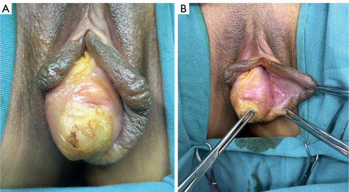
A vaginal mass located on the left vaginal wall, positioned beneath the mucosa near the vaginal orifice (A,B).
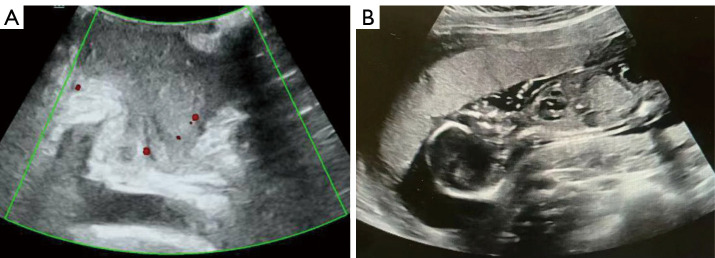
Ultrasound imaging indicated a vaginal mass measuring approximately 5 cm (A) and an intrauterine pregnancy (B).
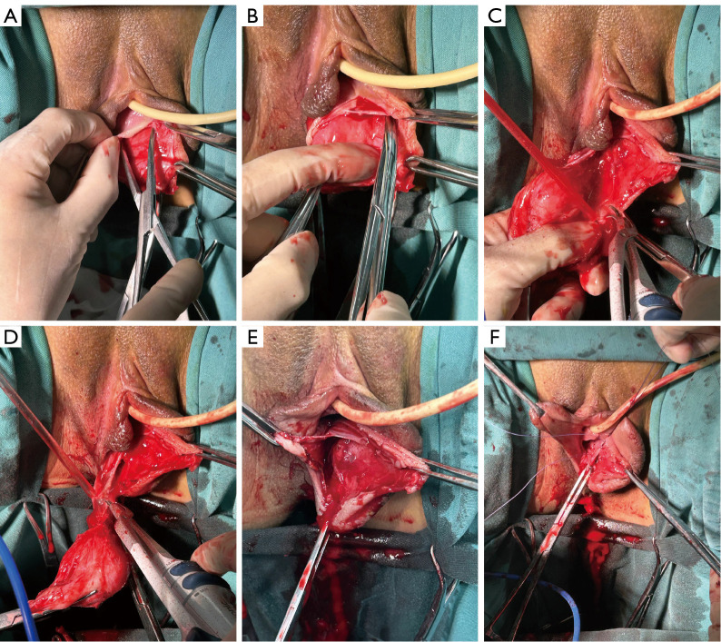
The steps of the operation. (A) The procedure began with an incision in the vaginal mucosa. (B) Scissors were used to carefully separate the mass from the vaginal wall. (C,D) An ultrasound knife was employed to cut the vessels supplying the mass. (E) The mass was completely resected, and the wound surface showed no obvious bleeding. (F) The vaginal mucosa was sutured with absorbable stitches.
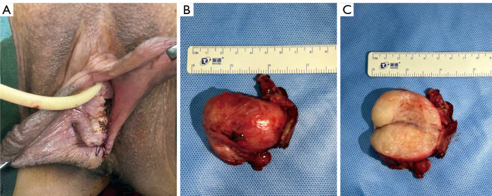
The appearance of the wound after the operation (A) and the size of the mass (B,C).
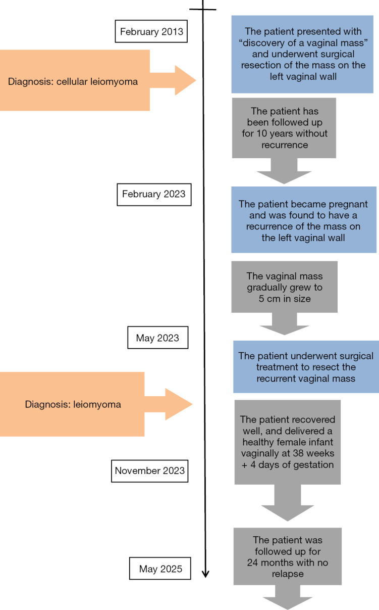
A timeline summarizing the patient’s clinical course.
Similar articles
-
Urethral Leiomyoma in Pregnancy: A Rare Case of Symptomatic Pelvic Mass With Successful Surgical Management.Cureus. 2025 Jun 25;17(6):e86720. doi: 10.7759/cureus.86720. eCollection 2025 Jun. Cureus. 2025. PMID: 40718265 Free PMC article.
-
Impact of definitive uterine artery occlusion on ovarian reserve markers in laparoscopic myomectomy: a randomized controlled trial with 2-year follow-up.Hum Reprod. 2025 Jul 1;40(7):1305-1314. doi: 10.1093/humrep/deaf070. Hum Reprod. 2025. PMID: 40420404 Free PMC article. Clinical Trial.
-
Abdominal wound dehiscence after appendectomy during pregnancy treated by negative pressure wound therapy with subsequent vaginal delivery: A case report and literature review.Int J Gynaecol Obstet. 2025 Jul;170(1):150-156. doi: 10.1002/ijgo.16155. Epub 2025 Jan 18. Int J Gynaecol Obstet. 2025. PMID: 39825682 Free PMC article. Review.
-
Diagnosis and management dilemma in leiomyoma of the anterior vaginal wall.BMJ Case Rep. 2024 Dec 20;17(12):e262747. doi: 10.1136/bcr-2024-262747. BMJ Case Rep. 2024. PMID: 39950656
-
Progestogens for preventing miscarriage: a network meta-analysis.Cochrane Database Syst Rev. 2021 Apr 19;4(4):CD013792. doi: 10.1002/14651858.CD013792.pub2. Cochrane Database Syst Rev. 2021. PMID: 33872382 Free PMC article.
References
-
- Shrestha A, Mudbari J, Tamrakar SR, Pradhan N, Makaju R, Karki S. A Rare Case of Large Left Lateral Wall Vaginal Myoma. Kathmandu Univ Med J (KUMJ) 2021;19:396-8. - PubMed
-
- Dane C, Rustemoglu Y, Kiray M, Ozkuvanci U, Tatar Z, Dane B. Vaginal leiomyoma in pregnancy presenting as a prolapsed vaginal mass. Hong Kong Med J 2012;18:533-5. - PubMed
Publication types
LinkOut - more resources
Full Text Sources