

Incidence, outcome, and risk factors of contrast media extravasation injury during contrast-enhanced computed tomography scans: an observational cohort study
- PMID: 40785883
- PMCID: PMC12332564
- DOI: 10.21037/qims-24-2332
Incidence, outcome, and risk factors of contrast media extravasation injury during contrast-enhanced computed tomography scans: an observational cohort study
Abstract
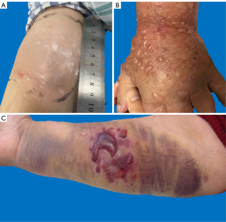
Background: Contrast media (CM) is widely used in contrast-enhanced computed tomography (CECT) to enhance the visualization of abnormal structures. CM extravasation, a recognized complication, may cause mild swelling or severe injuries (e.g., ulceration, necrosis). Current research on severe extravasation is limited to case reports or lacks large-cohort analyses of risk factors. This study aimed to explore risk factors, validate incidence and outcomes, and compare patient/technique/CM-related factors between mild and severe CM extravasation injuries using large-scale clinical data to aid early recognition and prevention.
Methods: This is a retrospective cohort study. A total of 586,812 CM injections were performed during CECT scans at a single institution between November 2012 and December 2023. Among these, 709 cases (334 males, 375 females; age: 62.9±15.2 years) with CM extravasation injuries were included. Extravasation injuries were classified by severity. The frequency and clinical outcomes of different severities of injuries were investigated. Risk factors of serious injuries were evaluated using logistic regression with generalized estimating equation analyses.
Results: CM extravasation occurred in 0.12% (709/586,812) of cases. Of the 709 extravasation injuries, 672 (94.8%) were mild, 32 (4.5%) were moderate, and 5 (0.7%) were severe. Only 5 patients received consultations from dermatologists or burn specialists, and none required surgery. Multivariate analysis underscored the presence of diabetes mellitus [DM, odds ratio (OR) =8.04; P<0.01], injections in the dorsum of the hand (OR =4.86; P<0.01), without saline test (OR =2.58; P=0.02), and large-volume extravasation (OR =5.49; P<0.01) as potential risk factors of moderate or severe CM extravasation injury.
Conclusions: Most CM extravasation injuries are mild and without serious consequences. Multiple modifiable risk factors for serious CM extravasation injury have been identified that could mitigate the severity of the injury.
Keywords: Contrast media (CM); computed tomography (CT); extravasation of diagnostic and therapeutic materials; injection site reactions.
Copyright © 2025 AME Publishing Company. All rights reserved.
Conflict of interest statement
Conflicts of Interest: All authors have completed the ICMJE uniform disclosure form (available at https://qims.amegroups.com/article/view/10.21037/qims-24-2332/coif). The authors have no conflicts of interest to declare.
Figures
Similar articles
-
Contrast-enhanced ultrasound using SonoVue® (sulphur hexafluoride microbubbles) compared with contrast-enhanced computed tomography and contrast-enhanced magnetic resonance imaging for the characterisation of focal liver lesions and detection of liver metastases: a systematic review and cost-effectiveness analysis.Health Technol Assess. 2013 Apr;17(16):1-243. doi: 10.3310/hta17160. Health Technol Assess. 2013. PMID: 23611316 Free PMC article.
-
Comparison of Two Modern Survival Prediction Tools, SORG-MLA and METSSS, in Patients With Symptomatic Long-bone Metastases Who Underwent Local Treatment With Surgery Followed by Radiotherapy and With Radiotherapy Alone.Clin Orthop Relat Res. 2024 Dec 1;482(12):2193-2208. doi: 10.1097/CORR.0000000000003185. Epub 2024 Jul 23. Clin Orthop Relat Res. 2024. PMID: 39051924
-
Systemic pharmacological treatments for chronic plaque psoriasis: a network meta-analysis.Cochrane Database Syst Rev. 2021 Apr 19;4(4):CD011535. doi: 10.1002/14651858.CD011535.pub4. Cochrane Database Syst Rev. 2021. Update in: Cochrane Database Syst Rev. 2022 May 23;5:CD011535. doi: 10.1002/14651858.CD011535.pub5. PMID: 33871055 Free PMC article. Updated.
-
Drugs for preventing postoperative nausea and vomiting in adults after general anaesthesia: a network meta-analysis.Cochrane Database Syst Rev. 2020 Oct 19;10(10):CD012859. doi: 10.1002/14651858.CD012859.pub2. Cochrane Database Syst Rev. 2020. PMID: 33075160 Free PMC article.
-
Sexual Harassment and Prevention Training.2024 Mar 29. In: StatPearls [Internet]. Treasure Island (FL): StatPearls Publishing; 2025 Jan–. 2024 Mar 29. In: StatPearls [Internet]. Treasure Island (FL): StatPearls Publishing; 2025 Jan–. PMID: 36508513 Free Books & Documents.
References
-
- Wu TL, Chang TY, Su TC, Huo Teng M, Hsu HC. Acute adverse reactions and extravasation of iodinated contrast media during computerized tomography scans: A 7-year experience in a local teaching hospital. J Radiol Sci 2023;48:13.
-
- Cariaga R. Standardization of access for administration of intravenous contrast for computed tomography scans. Standardization of PIV Line Preparation for CT Scan 2021 May [cited 2024 Oct 9]; Available online: https://archive.hshsl.umaryland.edu/handle/10713/15691
LinkOut - more resources
Full Text Sources