

Clinical and imaging characteristics of toxoplasmic encephalitis in patients with HIV/AIDS with different immune statuses
- PMID: 40785890
- PMCID: PMC12332595
- DOI: 10.21037/qims-2024-2598
Clinical and imaging characteristics of toxoplasmic encephalitis in patients with HIV/AIDS with different immune statuses
Abstract
Background: Toxoplasmic encephalitis (TE) is one of the most common opportunistic central nervous system infections in patients with acquired immunodeficiency syndrome (AIDS) and a major cause of death. Magnetic resonance imaging (MRI) serves as a key adjunct in the clinical diagnosis of TE. This study aimed to identify the MRI characteristics of TE in patients with human immunodeficiency virus (HIV)/AIDS across different immune statuses.
Methods: A retrospective analysis was conducted on the clinical and imaging data of hospitalized patients with AIDS and concurrent TE treated at two centers between January 2019 and December 2023. Patients were divided into three groups based on CD4+ T-cell count levels (<50, 50-100, and >100 cells/µL). The differences in MRI imaging features between patients with varying immune statuses were analyzed.
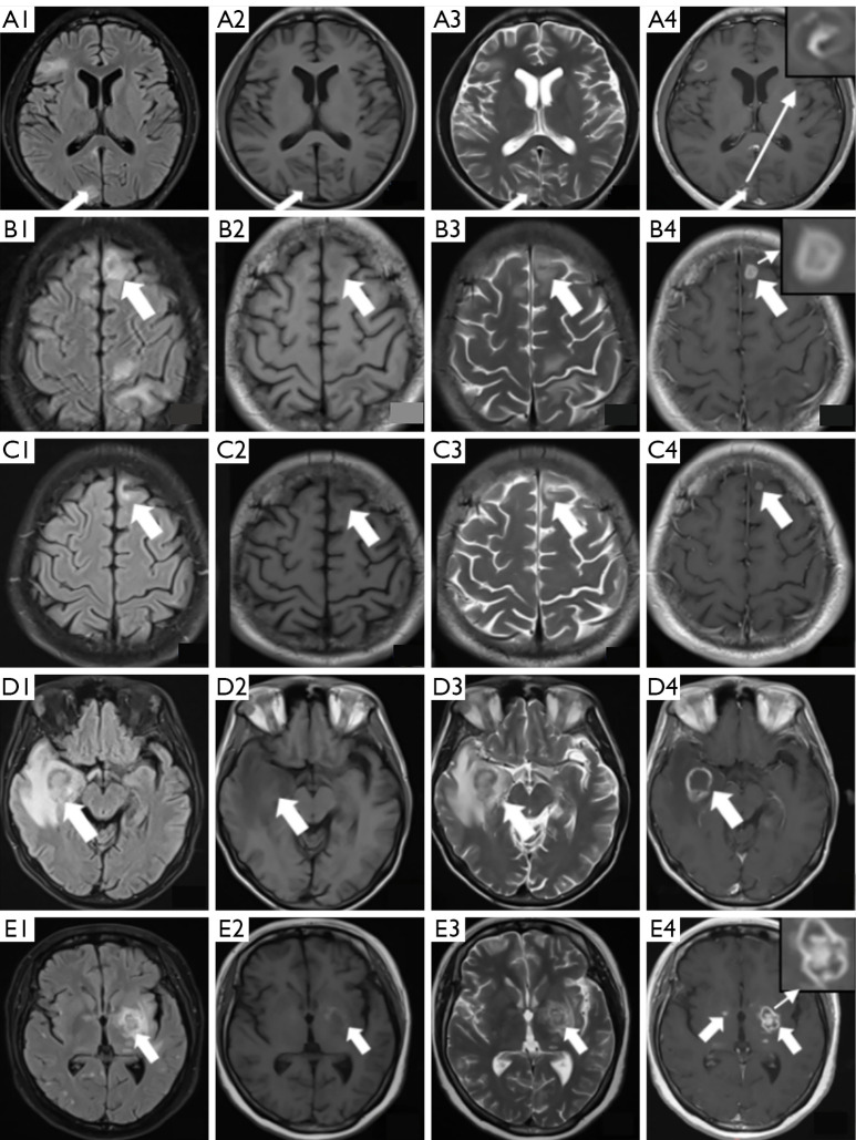
Results: A total of 41 patients were included, 82.9% had multiple lesions on MRI, and 203 lesions larger than 2 mm were identified. The lesions predominantly presented with nodular and ring-enhancing patterns, mainly distributed at the gray-white matter junction and the thalamus-basal ganglia region. In the comparison of MRI features between the three groups, patients with >100 CD4+ T cells/µL had a higher prevalence of nodular lesions and eccentric target lesions (both P<0.05). For patients with <50 CD4+ T cells/µL, larger lesions (>2 cm) were more frequently observed on MRI (P<0.05). Additionally, the <50 CD4+ T cells/µL group showed significantly elevated cerebrospinal fluid protein levels as compared to the other two groups (P<0.05).
Conclusions: In patients with HIV/AIDS with secondary TE, MRI findings varied in terms of lesion size and enhancement patterns according to the immune status of the patient. These imaging characteristics provide valuable guidance for the clinical diagnosis and treatment of TE.
Keywords: Toxoplasmosis; acquired immunodeficiency syndrome (AIDS); brain; human immunodeficiency virus (HIV); magnetic resonance imaging (MRI).
Copyright © 2025 AME Publishing Company. All rights reserved.
Conflict of interest statement
Conflicts of Interest: All authors have completed the ICMJE uniform disclosure form (available at https://qims.amegroups.com/article/view/10.21037/qims-2024-2598/coif). The authors have no conflicts of interest to declare.
Figures
References
-
- Lewden C, Drabo YJ, Zannou DM, Maiga MY, Minta DK, Sow PS, Akakpo J, Dabis F, Eholié SP; IeDEA West Africa Collaboration. Disease patterns and causes of death of hospitalized HIV-positive adults in West Africa: a multicountry survey in the antiretroviral treatment era. J Int AIDS Soc 2014;17:18797. 10.7448/IAS.17.1.18797 - DOI - PMC - PubMed
-
- Okome-Nkoumou M, Guiyedi V, Ondounda M, Efire N, Clevenbergh P, Dibo M, Dzeing-Ella A. Opportunistic diseases in HIV-infected patients in Gabon following the administration of highly active antiretroviral therapy: a retrospective study. Am J Trop Med Hyg 2014;90:211-5. 10.4269/ajtmh.12-0780 - DOI - PMC - PubMed
LinkOut - more resources
Full Text Sources
Research Materials