Review
. 2025 Aug 5:20:9695-9721.
doi: 10.2147/IJN.S520205. eCollection 2025.

Research Progress on Nucleus-Targeting Carbon Nanoparticles for Tumor Imaging and Therapy

Affiliations
Review

Research Progress on Nucleus-Targeting Carbon Nanoparticles for Tumor Imaging and Therapy

Hongkai Mu et al. Int J Nanomedicine. .

Abstract

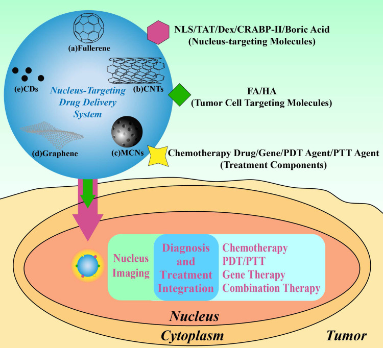
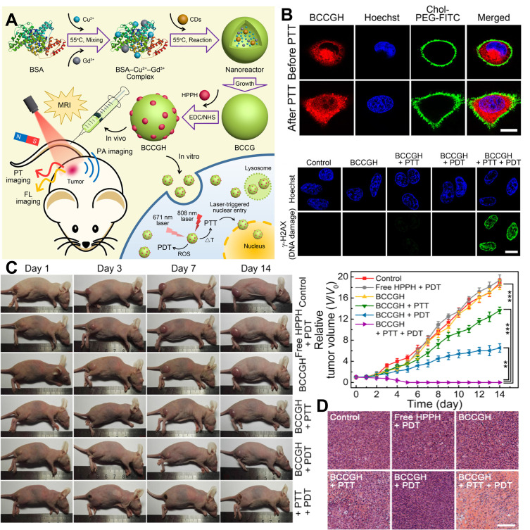
The cell nucleus, as the central hub of cellular physiological activities, represents an effective target for cancer therapy. Targeting strategies aimed at the cell nucleus can enhance the delivery of more drugs into the tumor cell nucleus, thereby improving therapeutic efficacy. Such approaches can overcome the limitations of previous tissue- and cell-level treatments, such as insufficient specificity and excessive toxic side effects. Currently, researchers have integrated carbon nanoparticles (CNPs), therapeutic molecules, and nucleus-targeting components to construct various nucleus-targeted carbon nanoparticle delivery systems for tumor imaging and treatment. These systems not only increase drug concentration within the cell nucleus but also enable certain CNPs to utilize their intrinsic fluorescence for imaging monitoring. This review summarizes the latest classification progress of nucleus-targeted CNPs, their mechanisms of action in cancer-targeted diagnosis and treatment, and their applications in cancer therapy. Additionally, the article discusses the current challenges and future directions in this field, aiming to provide valuable reference information for precision cancer treatment.

Keywords: carbon nanoparticles; nucleus-targeting; tumor imaging and therapy.


Conflict of interest statement

The authors declare that they have no known competing financial interests or personal relationships that could have appeared to influence the work reported in this paper.

Figures

Figure 1
Figure 1
Strategies for construction of nucleus-targeting drug delivery system based on CNPs for imaging and therapy.
Figure 2
Figure 2
The targeting nucleus mechanism of NPs.
Figure 3
Figure 3
Schematic representation of NPs translocating into the nucleus through NLS.
Figure 4
Figure 4
Synthesis route of nucleus-targeting CNPs. (A) Synthesis route of C60-serPF; reproduced from Raoof M, Mackeyev Y, Cheney MA, Wilson LJ, Curley SA. Internalization of C60 fullerenes into cancer cells with accumulation in the nucleus via the nuclear pore complex. Biomaterials. 2012;33:2952–2960. Copyright © 2011 Elsevier Ltd. All rights reserved. (B) Synthesis route of DOX/PGA-Hep-MWCNTs; Reproduced from Tsai H-C, Maryani F, Huang -C-C, Imae T, Lin J-Y. Drug-loading capacity and nuclear targeting of multiwalled carbon nanotubes grafted with anionic amphiphilic copolymers. Int J Nanomed. 2013;8:4427. © 2013 The Author(s). Dove Medical Press Limited. Creative Commons Attribution - Non Commercial (unported, 3.0) License. (C) Synthesis route of DOX/OX-MPCNP-Cys-PAsp-FA; Reproduced from Vinothini K, Dhilip Kumar SS, Abrahamse H, Rajan M. Enhanced doxorubicin delivery in folate-overexpressed breast cancer cells using mesoporous carbon nanospheres. ACS Omega. 2021;6:34532–34545. © 2021 The Authors. Published by American Chemical Society. https://creativecommons.org/licenses/by-nc-nd/4.0/. (D) Synthesis route of TG; Reproduced from Shan S, Jia S, Lawson T, Yan L, Lin M, Liu Y. The use of TAT peptide-functionalized graphene as a highly nuclear-targeting carrier system for suppression of choroidal melanoma. Int J Mol Sci. 2019;20:4454. © 2019 by the authors. Licensee MDPI, Basel, Switzerland. This article is an open access article distributed under the terms and conditions of the Creative Commons Attribution (CC BY) license. (E) Synthesis route of DOX/CQD; Reproduced from Jung YK, Shin E, Kim B-S. Cell nucleus-targeting zwitterionic carbon dots. Sci Rep. 2015;5. Copyright © 2015, Macmillan Publishers Limited. This work is licensed under a Creative Commons Attribution 4.0 International License. (F) Synthesis route of GQD/DOX; Reproduced from Wang C, Wu C, Zhou X, et al. Enhancing cell nucleus accumulation and DNA cleavage activity of anti-cancer drug via graphene quantum dots. Sci Rep. 2013;3. Copyright © 2013, The Author(s). Creative Commons CC-BY-NC-ND license.
Figure 5
Figure 5
Images for nucleus imaging using nucleus-targeting CNPs: (A) CLSM images of B16-F10 cells and TAT-CDs; Reproduced from Song Y, Li X, Cong S, Zhao H, Tan M. Nuclear-targeted of TAT peptide-conjugated carbon dots for both one-and two-photon fluorescence imaging. © 2019 Elsevier B.V. All rights reserved. (B) CLSM image of NLS-CDs incubated with MCF7 cells for 4 h; Repproduced from Yang L, Jiang W, Qiu L, et al. One pot synthesis of highly luminescent polyethylene glycol anchored carbon dots functionalized with a nuclear localization signal peptide for cell nucleus imaging. Nanoscale. 2015;7:6104–6113. Copyright 2019, Royal Society of Chemistry. (C) CLSM images of HeLa cells incubated with CDs; Reproduced from Yin X, Sun Y, Yang R, Qu L, Li Z. RNA-responsive fluorescent carbon dots for fast and wash-free nucleolus imaging, Spectrochim. Acta A Mol Biomol Spectrosc. 2020;237:118381. © 2020 Elsevier B.V. All rights reserved. (D) The localization of Ni-pPCDs and SYTORNASelect in cells; Reproduced from Hua X-W, Bao Y-W, Zeng J, Wu F-G. Nucleolus-targeted red emissive carbon dots with polarity-sensitive and excitation-independent fluorescence emission: high-resolution cell imaging and in vivo tracking. ACS Appl Mater Interfaces. 2019;11:32647–32658. Copyright © 2019 American Chemical Society. (E) Asynchronous cell imaging and (F) polychromatic laser CLSM for HeLa cells; Reproduced from Zhang Y, Zhang X, Shi Y, Sun C, Zhou N, Wen H. The synthesis and functional study of multicolor nitrogen-doped carbon dots for live cell nuclear imaging. Molecules. 2020;25:306. © 2020 by the authors. Licensee MDPI, Basel, Switzerland. This article is an open access article distributed under the terms and conditions of the Creative Commons Attribution (CC BY) license. (G) CLSM images of HeLa cells and HBIE-CDs; Reproduced from Liu H, Yang J, Li Z, et al. Hydrogen-bond-induced emission of carbon dots for wash-free nucleus imaging. Analy Chem. 2019;91:9259–9265. Copyright © 2019 American Chemical Society.
Figure 6
Figure 6
Images for chemotherapy using nucleus-targeting CNPs: (A) CLSM image of Dox@h-FCD with HeLa cells; (B) Viability of HeLa cells in different groups; (C) CLSM of MCF7 cells incubated with experimental group; (D) The condition of tumors post-treatment with materials from different groups; (E) Photographs of mouse tumors taken at different time points after treatment in the experimental group, as well as ex vivo tumor images from different groups following treatment; Das M, Singh RP, Datir SR, Jain S. Intranuclear drug delivery and effective in vivo cancer therapy via estradiol–peg-appended multiwalled carbon nanotubes. Mol Pharm. 2013;10:3404–3416. Copyright © 2013 American Chemical Society. (F) Images of MCF7/HER2 cells incubated with experimental group; (G) Viability of MCF7/HER2 cells in different groups; Reproduced from Zheng XT, Ma XQ, Li CM. Highly efficient nuclear delivery of anti-cancer drugs using a bio-functionalized reduced graphene oxide. J Colloid Interf Sci. 2016;467:35–42. Copyright © 2016 Elsevier Inc. All rights reserved. (H) Tumor conditions following the administration of different group treatments; (I) CLSM of CSCNP-R-CQDs incubated with HeLa cells and Breast CSCs; Reproduced from Su W, Guo R, Yuan F, et al. Red-emissive carbon quantum dots for nuclear drug delivery in cancer stem cells. J Phys Chem Lett. 2020;11:1357–1363. Copyright © 2020 American Chemical Society. (J) CLSM images of HepG2 cells incubated with CDs; (K) Tumor suppression curves for CDs and CDs/GA groups; Reproduced from Liu S, Wang J, Li H, et al. Nucleus‐targeting carbon quantum dots assembled with gambogic acid via π–π stacking for cancer therapy. Adv Ther. 2022;6. © 2022 Wiley‐VCH GmbH. ***p < 0.001, ****p < 0.0001.
Figure 7
Figure 7
Images for PTT/PDT using nucleus-targeting CNPs: (A) The synthesis of BCCGH and its applications in PTT and PDT; (B) CLSM of A549 cells before and after PTT treatment, as well as CLSM of A549 cells following the various treatments indicated; (C) Photographs of tumor-bearing mice after the indicated treatment and a summary of tumor growth in each group. Reproduced from Hua XW, Bao YW, Zeng J, Wu FG. Ultrasmall all-in-one nanodots formed via carbon dot-mediated and albumin-based synthesis: multimodal imaging-guided and mild laser-enhanced cancer therapy. ACS Appl Mater Interfaces. 2018;10:42077–42087. Copyright © 2018 American Chemical Society. **P < 0.01, ***P < 0.001; (D) H&E stained sections of tumor tissue.
Figure 8
Figure 8
Images for gene therapy using nucleus-targeting CNPs: (A) Schematics representation of CQDs an effective tool for genome editing and fluorpDNA was taken up by cells through caveolae-mediated endocytosis. Caveosomes containing CQDsininpDNA traffic to the Golgi and/or endoplasmic reticulum (ER), which then penetrate into the cytosol, ultimately enter the nucleus via nuclear pore complexes through passive diffusion. CQDs–P/pDNA was taken up by cells through clathrin-mediated endocytosis. Endocytic vesicles would deliver CQDsivepDNA to lysosomes and thus limit their release. Even if CQDsn ipDNA escape from lysosomes, they cannot enter the nucleus through NPCs due to exceeding the threshold diameter for passive diffusion; (B) Confocal laser scanning microscopy images of pX459-EFHD1-sgRNA transfected into HeLa cells for 45 minutes; (C) Genomic DNA sequencing profile; (D) Indel spectrum following puromycin resistance selection. Reproduced from Zhai LM, Zhao Y, Xiao RL, et al. Nuclear-targeted carbon quantum dot mediated CRISPR/Cas9 delivery for fluorescence visualization and efficient editing. Nanoscale. 2022;14:14645–14660. Copyright 2022 Royal Society of Chemistry.
Figure 9
Figure 9
Images for combination therapy using nucleus-targeting CNPs: (A) The synthesis and synergistic therapeutic mechanism of HKHD; (B) CLSM images of A549 cells treated with HKHD; (C) The growth and inhibition of tumors following different treatments are shown. *P < 0.05; **P < 0.01; (D) H&E staining was used to evaluate tumor apoptosis. Reproduced from Xie R, Lian S, Peng H, et al. Mitochondria and nuclei dual-targeted hollow carbon nanospheres for cancer chemophotodynamic synergistic therapy. Mol Pharm. 2019;16:2235–2248. Copyright © 2019 American Chemical Society.
Figure 10
Figure 10
Images for diagnosis and treatment using nucleus-targeting CNPs: (A) Schematic illustration of the FRET system based on GQDs for nucleus-targeting drug delivery; (B) Quantitative detection of DOX after incubation of cells with the indicated materials; (C) Survival rate of HeLa cells incubated with the indicated materials; (D) Changes in FRET signals following treatment with the indicated materials; Reproduced from Chen HS, Wang Z, Zong S, et al. A graphene quantum dot-based FRET system for nuclear-targeted and real-time monitoring of drug delivery. Nanoscale. 2015;7:15477–15486. Copyright 2015 The Royal Society of Chemistry. (E) CLSM images at different time points after treatment with CD-PpIX, and the corresponding fluorescence-intensity-profile analysis of the marked arrows in (E). Red line: PpIX, blue line: Hoechst 33342; (F) Tumor growth curves in mice after the indicated treatments; Reproduced from Hua XW, Bao YW, Wu FG. Fluorescent carbon quantum dots with intrinsic nucleolus-targeting capability for nucleolus imaging and enhanced cytosolic and nuclear drug delivery. ACS Appl Mater Interfaces. 2018;10:10664–10677. Copyright © 2018 American Chemical Society. ***P < 0.001.

Similar articles

References

    1. Siegel RL, Miller KD, Wagle NS, Jemal A. Cancer statistics, 2023. CA. 2023;73:17–48. doi: 10.3322/caac.21763 - DOI - PubMed
    1. Damia G, Garattini S. The pharmacological point of view of resistance to therapy in tumors. Cancer Treat Rev. 2014;40:909–916. doi: 10.1016/j.ctrv.2014.05.008 - DOI - PubMed
    1. Raju B, Choudhary S, Narendra G, Verma H, Silakari O. Molecular modeling approaches to address drug-metabolizing enzymes (DMEs) mediated chemoresistance: a review. Drug Metab Rev. 2021;53:45–75. doi: 10.1080/03602532.2021.1874406 - DOI - PubMed
    1. Lin Z, Wu Y, Xu Y, Li G, Li Z, Liu T. Mesenchymal stem cell-derived exosomes in cancer therapy resistance: recent advances and therapeutic potential. Mol Cancer. 2022;21. doi: 10.1186/s12943-022-01650-5 - DOI - PMC - PubMed
    1. Ramirez JAZ, Romagnoli GG, Kaneno R. Inhibiting autophagy to prevent drug resistance and improve anti-tumor therapy. Life Sci. 2021;265:118745. doi: 10.1016/j.lfs.2020.118745 - DOI - PubMed

LinkOut - more resources