Review
. 2025 Aug 8:8:0816.
doi: 10.34133/research.0816. eCollection 2025.

Deciphering Mitochondria: Unveiling Their Roles in Mechanosensing and Mechanotransduction

Affiliations
Review

Deciphering Mitochondria: Unveiling Their Roles in Mechanosensing and Mechanotransduction

Jiaxuan Yu et al. Research (Wash D C). .

Abstract

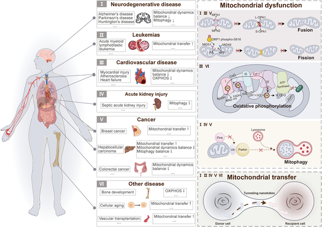
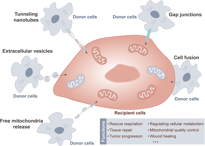
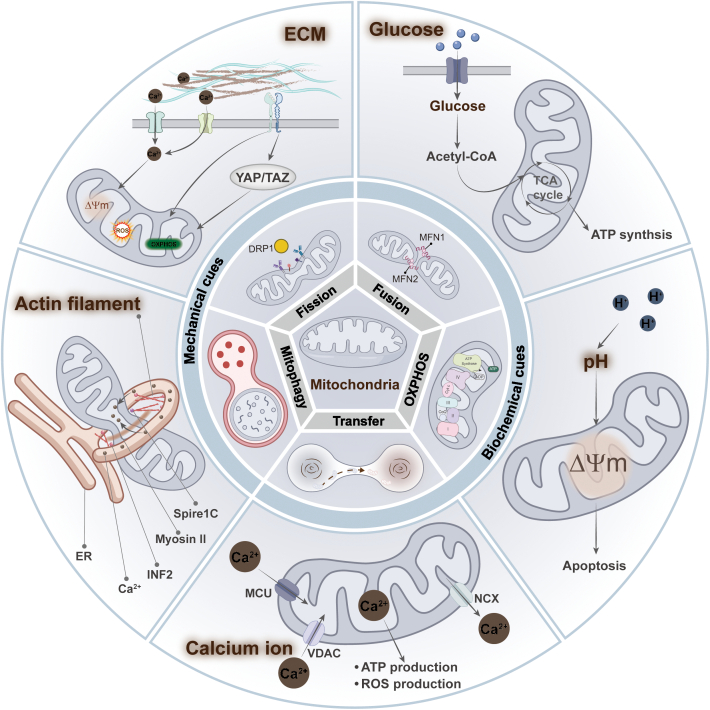
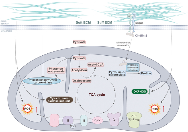
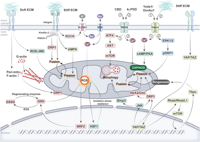
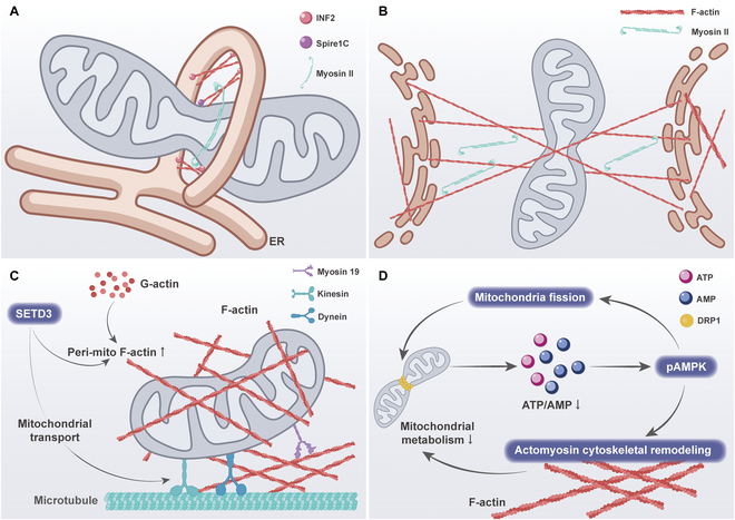
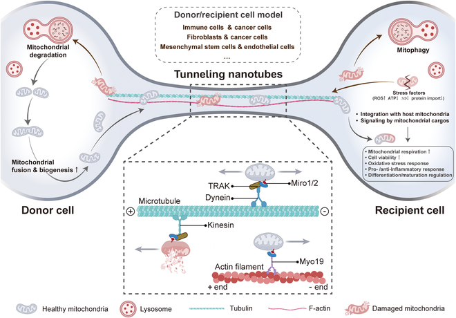
Mitochondria are highly dynamic organelles that are responsible for essential cellular functions such as calcium regulation, reactive oxygen species (ROS) production, metabolism, and apoptosis initiation. Mitochondrial dysfunctions are associated with a variety of pathologies, and the onset and progression of disease are accompanied by alterations in extracellular biochemical and mechanical signals. Recent studies have demonstrated that physicochemical cues, especially mechanical cues, exert pivotal roles in the organization of mitochondrial network and their metabolic functions. Therefore, understanding the mechanisms that orchestrate mitochondrial morphology and function is essential for elucidating their role in both health and disease. This review discusses novel insights into the recent advances regarding mitochondrial dysfunction across a spectrum of diseases and describes the effect of various factors. It then highlights the recently discovered mechanisms, particularly those involving matrix mechanical cues and cellular mechanical cues, summarizing the multiple pathways of mechanotransduction, such as integrin, Piezo1/TRPV4, and YAP/TAZ signaling pathways. Last, the review explores the potential future directions, stressing that understanding mitochondrial dysfunction is crucial for developing effective therapies to improve mitochondrial function and address related diseases.


Conflict of interest statement

Competing interests: The authors declare that they have no competing interests.

Figures

Fig. 1.
Fig. 1.
Dysfunctional mitochondria and diseases. Mitochondrial dysfunction includes imbalances in mitochondrial dynamics, impaired mitochondrial OXPHOS, and abnormal mitophagy. In addition, mitochondria can be transferred to other cells via TNTs. Mitochondrial dysfunction has been associated with a variety of diseases characterized by different mitochondrial dysfunctions. I: Neurodegenerative diseases, such as Alzheimer’s, Parkinson’s, and Huntington’s, have been associated with an imbalance in mitochondrial dynamics and a decrease in mitophagy. II: Leukemias, such as AML, have been associated with an increase in intercellular mitochondrial transfer. III: CVDs such as myocardial injury, atherosclerosis, and HF are associated with impaired OXPHOS and imbalanced mitochondrial dynamics. IV: AKI has been associated with reduced mitophagy. V: Cancers such as breast cancer, hepatocellular carcinoma, and colorectal cancer display mitochondrial dysfunction. These defects involve pathological alterations in mitochondrial transfer, dynamics, and mitophagy. VI: Other diseases, such as bone development, aging, and vascular transplantation, show impaired OXPHOS and increased mitochondrial transfer.
Fig. 2.
Fig. 2.
Common modes and routes of mitochondrial transfer. Mitochondrial transfer can be achieved by intercellular contacts such as cell fusion, gap junctions, and TNTs. In addition, mitochondria can be translocated into other cells via extracellular vesicles or free mitochondrial release. The transfer of mitochondria from donor cells to recipient cells is closely related to rescue respiration, tissue repair, and tumor progression.
Fig. 3.
Fig. 3.
Biochemical and mechanical cues reorganize and regulate mitochondrial networks and functions. Biochemical cues include extracellular glucose concentration, pH, and Ca2+ concentration. Glucose concentration can regulate mitochondrial OXPHOS and ATP production by affecting the tricarboxylic acid (TCA) cycle. Changes in pH can induce alterations in mitochondrial membrane potential, triggering apoptosis via the mitochondrial pathway. On the one hand, Ca2+ can promote ATP production. On the other hand, Ca2+ overloaded in mitochondria may lead to elevated ROS levels. Mechanical cues include the physical properties of the ECM and intracellular cytoskeleton dynamics. ECM stiffness cause changes in mitochondrial dynamics and function by transmitting signals into the cell through mechanosensitive receptors on the cell membrane. The actomyosin network, in turn, is involved in regulating mitochondrial division processes, which indirectly affect mitochondrial function.
Fig. 4.
Fig. 4.
The relationship between ECM stiffness and mitochondrial metabolism. Softer matrix stiffness induced an increase in mitochondrial ROS levels and decrease in ATP levels in breast cancer cells. Mitochondrial PCK2 expression increased with increasing matrix stiffness, which activated glycolysis to promote osteogenesis in MSCs. In lung adenocarcinoma, stiffening of the ECM promotes translocation of kindlin-2 to mitochondria and interaction with the enzyme PYCR1, which induces an increase in proline synthesis and cellular proliferation in tumor cells. In human mammary epithelial cells, the morphology of mitochondria gradually changes as the matrix stiffness increases, which leads to increased intracellular ROS levels and decreased ATP levels. During the spreading of MSCs on the stiff matrix, there is an increase in mitochondrial division and decrease in ATP levels, which increases osteogenic differentiation of MSCs. In HCC cells, mitochondria were fragmented and granular in the softer environment, with down-regulation of mitochondria-encoded cytochrome c oxidase I, which inhibited cell proliferation.
Fig. 5.
Fig. 5.
Integration of mechanotransduction pathways and mitochondrial dynamics and function. Mechanotransduction pathways can be divided into 3 categories: the integrin pathway, the Piezo1/TRPV4 pathway, and the YAP/TAZ pathway. Cells sense alterations in ECM stiffness through integrin receptors. The soft matrix promotes mitochondrial division by polymerization of actin around mitochondria. This activates the ROS–NRF2 pathway and promotes glutathione production to counteract oxidative stress. Soft matrix also promotes mitochondrial division by activating the ROS–JNK pathway. On the other hand, hard matrix regulates mitochondrial fusion and division by interacting with kindlin-2 and PINCH-1 of integrins, or via AMPK. The hard matrix also regulates the activity of SLC9A1 and NCX by the integrin–ROCK pathway, which overloads mitochondrial calcium ions and activates the ROS–HSF1 pathway. HSF1 activates the expression of genes related to antioxidative stress, which helps the cells to fight against oxidative stress. Piezo1 and TRPV4 are classes of mechanosensitive ion channels. The activation of TRPV4 allows the influx of calcium ions into the cell. This can promote mitochondrial OXPHOS and also trigger mitochondrial autophagy through the ATF4/AKT/mTOR pathway. Similarly, activation of Piezo1 also allows calcium ion influx, which, on one hand, promotes OXPHOS via the cAMP/PKA pathway and, on the other hand, induces DRP1-mediated mitochondrial division via the ERK1/2 pathway. YAP/TAZ is a class of mechanosensitive transcription factors. Activated YAP enters the nucleus and affects mitochondrial division by regulating DRP1 activity, which may also regulate mitochondrial autophagy via JNK/Bnip3. In addition, activated TAZ enters the nucleus and regulates mitochondrial biogenesis through the Rheb/RhebL1–mTOR–Tfam pathway.
Fig. 6.
Fig. 6.
The multiple links between actin and mitochondria. (A) Spire1C specifically localizes to mitochondria and promotes actin assembly around mitochondria by directly binding to and stimulating INF2 on the visceral ER. This leads to an increase in mitochondria–ER contact sites and an increase in mitochondrial fission. In particular, myosin 19 tethers mitochondria to ER-associated actin, enhancing ER–mitochondrial contact and promoting mitochondrial fission. (B) The contractile activity of myosin II leads to random, inhomogeneous deformation of the cytoskeletal network. Mitochondria are locally compressed between taut actin filaments, which result in localized invagination of the mitochondrial surface. This particular network structure may exert tangential forces on mitochondria to promote their division. (C) SETD3, located on the outer mitochondrial membrane, regulates the formation of periplasmic F-actin, which is essential for the maintenance of mitochondrial morphology, movement, and function. (D) Altered mitochondrial metabolic capacity causes changes in the intracellular ATP/AMP ratio, which in turn activates pAMPK. On the one hand, pAMPK regulates cytoskeletal dynamics by direct phosphorylation and inactivation of myosin. On the other hand, pAMPK sustains its continuous activation by regulating mitochondrial division.
Fig. 7.
Fig. 7.
Mechanotransduction in mitochondria transfer. Mitochondria can be transferred between cells via TNTs. This can occur between immune cells and cancer cells, fibroblasts and cancer cells, MSCs and ECs, and other homogeneous and heterogeneous cells. The donor cells transfer their healthy mitochondria via TNTs to the stimulated or damaged recipient cells. This process helps to restore the bioenergetic properties of the recipient cells, enhances cellular viability, reduces inflammatory processes, and promotes normalization of cellular functions. Similarly, damaged mitochondria that are transferred to donor cells are used for other mitochondrial fusion processes and mitochondrial biogenesis through degradation. Mitochondria exhibit 2 modes of movement in TNT: one is microtubule-driven transport and the other is actin-based transport. When mitochondria form a motor articulation complex through Miro1/2, TRAK, and dynein, it can move in both directions along microtubules. When bound to kinesin, it can only move along the plus end of the microtubule. In addition, mitochondria through Miro1/2 can also synergize with Myo19, which allows it to move along the minus end of actin.

Similar articles

  • Short-Term Memory Impairment.
    Cascella M, Al Khalili Y. Cascella M, et al. 2024 Jun 8. In: StatPearls [Internet]. Treasure Island (FL): StatPearls Publishing; 2025 Jan–. 2024 Jun 8. In: StatPearls [Internet]. Treasure Island (FL): StatPearls Publishing; 2025 Jan–. PMID: 31424720 Free Books & Documents.
  • From gene to mechanics: a comprehensive insight into the mechanobiology of LMNA mutations in cardiomyopathy.
    Veltrop RJA, Kukk MM, Topouzidou K, Didden L, Muchir A, van Steenbeek FG, Schurgers LJ, Harakalova M. Veltrop RJA, et al. Cell Commun Signal. 2024 Mar 27;22(1):197. doi: 10.1186/s12964-024-01546-5. Cell Commun Signal. 2024. PMID: 38539233 Free PMC article. Review.
  • Systemic Inflammatory Response Syndrome.
    Baddam S, Burns B. Baddam S, et al. 2025 Jun 20. In: StatPearls [Internet]. Treasure Island (FL): StatPearls Publishing; 2025 Jan–. 2025 Jun 20. In: StatPearls [Internet]. Treasure Island (FL): StatPearls Publishing; 2025 Jan–. PMID: 31613449 Free Books & Documents.
  • PIEZO1-mediated mechanotransduction regulates collagen synthesis on nanostructured 2D and 3D models of fibrosis.
    Rashidi N, Harasymowicz NS, Savadipour A, Steward N, Tang R, Oswald S, Guilak F. Rashidi N, et al. Acta Biomater. 2025 Jan 24;193:242-254. doi: 10.1016/j.actbio.2024.12.034. Epub 2024 Dec 13. Acta Biomater. 2025. PMID: 39675497
  • Management of urinary stones by experts in stone disease (ESD 2025).
    Papatsoris A, Geavlete B, Radavoi GD, Alameedee M, Almusafer M, Ather MH, Budia A, Cumpanas AA, Kiremi MC, Dellis A, Elhowairis M, Galán-Llopis JA, Geavlete P, Guimerà Garcia J, Isern B, Jinga V, Lopez JM, Mainez JA, Mitsogiannis I, Mora Christian J, Moussa M, Multescu R, Oguz Acar Y, Petkova K, Piñero A, Popov E, Ramos Cebrian M, Rascu S, Siener R, Sountoulides P, Stamatelou K, Syed J, Trinchieri A. Papatsoris A, et al. Arch Ital Urol Androl. 2025 Jun 30;97(2):14085. doi: 10.4081/aiua.2025.14085. Epub 2025 Jun 30. Arch Ital Urol Androl. 2025. PMID: 40583613 Review.

References

    1. Shen K, Pender CL, Bar-Ziv R, Zhang H, Wickham K, Willey E, Durieux J, Ahmad Q, Dillin A. Mitochondria as cellular and organismal signaling hubs. Annu Rev Cell Dev Biol. 2022;38:179–218. - PubMed
    1. Ni HM, Williams JA, Ding WX. Mitochondrial dynamics and mitochondrial quality control. Redox Biol. 2015;4:6–13. - PMC - PubMed
    1. Singh G, Vengayil V, Khanna A, Adhikary S, Laxman S. Active control of mitochondrial network morphology by metabolism-driven redox state. Proc Natl Acad Sci USA. 2025;122(16): Article e2421953122. - PMC - PubMed
    1. Chan DC. Mitochondrial dynamics and its involvement in disease. Annu Rev Pathol. 2020;15:235–259. - PubMed
    1. Chen W, Zhao H, Li Y. Mitochondrial dynamics in health and disease: Mechanisms and potential targets. Signal Transduct Target Ther. 2023;8(1):333. - PMC - PubMed

LinkOut - more resources