. 2025 Jul 25:16:1600349.
doi: 10.3389/fphar.2025.1600349. eCollection 2025.

A glycogen derived from sea urchin- Strongylocentyotus internedius shifts macrophages to the M1 phenotype and enhances the anti-pancreatic cancer activity of gemcitabine

Affiliations

A glycogen derived from sea urchin- Strongylocentyotus internedius shifts macrophages to the M1 phenotype and enhances the anti-pancreatic cancer activity of gemcitabine

Zhenzhen Deng et al. Front Pharmacol. .

Abstract

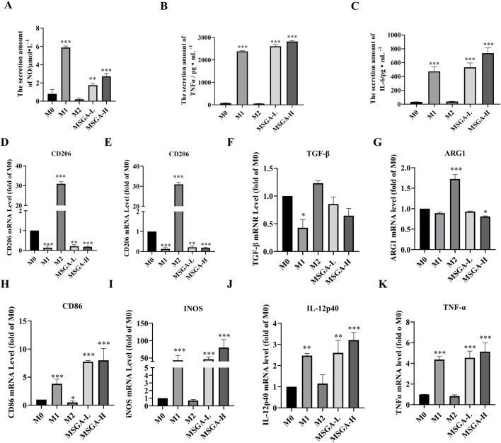
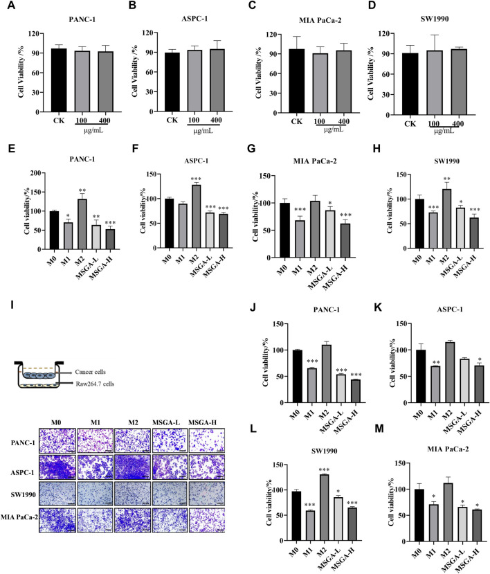
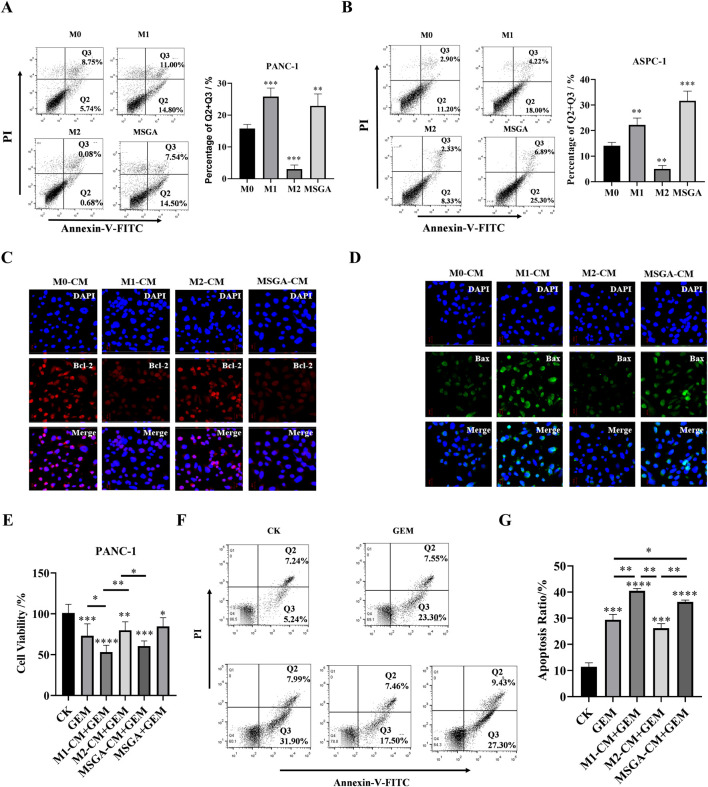
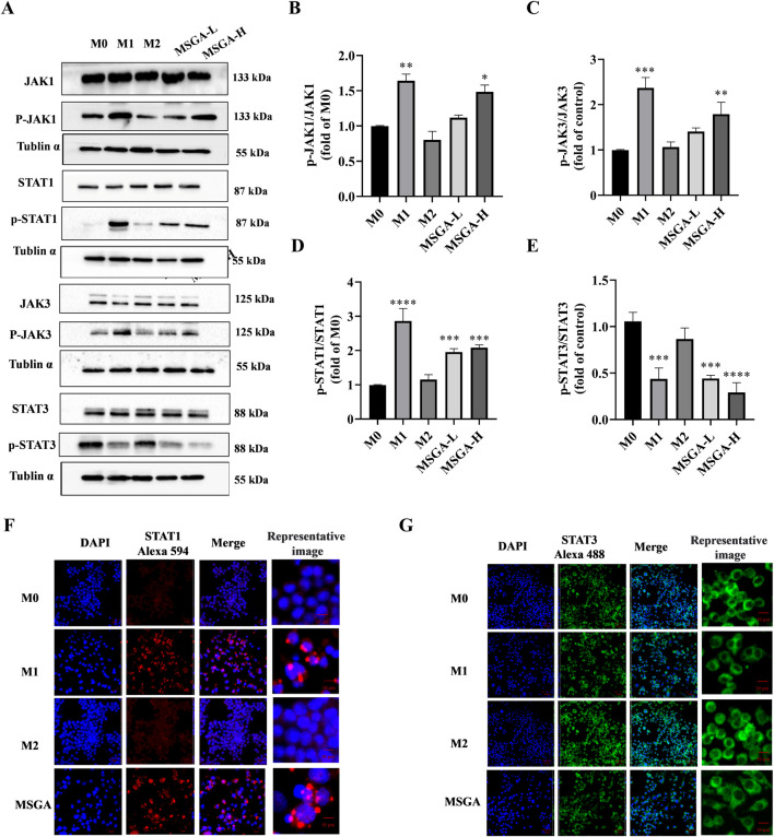
One of the biggest obstacles to treating pancreatic ductal adenocarcinoma (PDAC) is chemotherapy resistance. Macrophages are an essential element of the innate immune system and are distributed in almost every tissue in the body. Among them, macrophages infiltrating into the tumor microenvironment negatively regulate tumor immunity and participate in the generation, invasion, migration and drug resistance of PDAC. In prior study, we isolated a polysaccharide from sea urchin-Strongylocentyotus internedius, which was identified as a high molecular weight, highly branched glycogen (MSGA). In this study, we found that MSGA increased the expression of iNOS, IL-6, TNFα, IL-12 and triggered macrophage differentiation to the CD86+ M1 phenotype. MSGA-induced M1 macrophages decreased the cell viabilities and induced apoptosis of PDAC cells. When combined with gemcitabine (GEM), MSGA significantly enhanced the pro-apoptotic activity of GEM. Mechanistically, MSGA transformed macrophages to the M1 phenotype through the stimulation of the JAK1/3-STAT1 signaling pathway and the suppression of STAT3 activity. Overall, our research showed that MSGA has profound potential for tumor immunotherapy. And as an "immune stimulator", MSGA could assist GEM in the treatment of PDAC.

Keywords: JAK; STAT signaling pathway; gemcitabine; glycogen; macrophages; tumor microenvironment.


Conflict of interest statement

The authors declare that the research was conducted in the absence of any commercial or financial relationships that could be construed as a potential conflict of interest.

Figures

FIGURE 1
FIGURE 1
MSGA polarizes macrophages to CD86+ M1 phenotype. (A) Cell viability of Raw264.7 cells after the cells were treated with MSGA for 36 h, n = 6. (B) The morphology of Raw264.7 were observed and collected by scanning electron microscopy (SEM) after treated with MSGA, n = 3. Macrophages were labeled with CD86 and CD206, and flow cytometry and immunofluorescence (IF) were used to identify the phenotypic transformation of macrophage. The percentages of CD86+ or CD206+ Raw264.7 cells (C) or BMDMs (D), n = 3. Representative images captured by laser confocal after immunofluorescence labeling of macrophages with CD86 (E) and CD206 (F) (image scale, 20 μm), n = 3. MSGA-L (100 μg/mL); MSGA-H (400 μg/mL). Data were expressed as means ± SD. T-test and One-way ANOVA with Tukey’s post-hoc test were used for the statistical analyses. Compared with M0 macrophages, statistically significant differences are indicated. *p < 0.05, **p < 0.01 and ***p < 0.001.
FIGURE 2
FIGURE 2
MSGA increases the releases of inflammatory cytokines. The contents of NO (A), IL-6 (C) and TNFα (B) produced by MSGA-treated Raw264.7 cells were measured by ELISA, n = 6. The expressions of M1/M2 cytokines and surface markers in MSGA-treated macrophages were detected by real-time fluorescence quantitative PCR and expressed as a fold change compared with M0 macrophages, M2 biomarkers include CD206 (D), IL-10 (E), TGF-β (F) and ARG1 (G). M1 biomarkers include CD86 (H), INOS (I), IL-12 (J) and TNFα (K), n = 6. MSGA-L (100 μg/mL); MSGA-H (400 μg/mL). Data were expressed as means ± SD. One-way ANOVA with Tukey’s post-hoc test was used for the statistical analyses. Compared with M0 macrophages, statistically significant differences are indicated. * p < 0.05, ** p < 0.01 and *** p < 0.001.
FIGURE 3
FIGURE 3
MSGA inhibits the viability of PDAC cells by polarizing macrophages into an M1 phenotype. Cytotoxicity of PANC-1 (A), ASPC-1 (B), MIA-PaCa-2 (C) and SW1990 (D) after MSGA treated cells for 36 h. Cytotoxicity of PANC-1 (E), ASPC-1 (F), MIA-PaCa-2 (G) and SW1990 (H) after different CMs treated cancer cells for 36 h. (I). Different phenotypes of macrophages and MSGA pretreated macrophages were co-cultured with PDAC cells in transwell co-culture system. The representative images of the cancer cells were collected (scale bar, 100 μm), n = 3. The density of PANC-1 (J), ASPC-1 (K), SW1990 (L) and MIA-PaCa-2 (M) in upper chamber of transwell was analyzed by using Image (J). MSGA-L (100 μg/mL); MSGA-H (400 μg/mL). One-way ANOVA with Tukey’s post hoc test was used for the statistical analyses. Compared with M0 macrophages, statistically significant differences are indicated. * p < 0.05, ** p < 0.01 and *** p < 0.001.
FIGURE 4
FIGURE 4
MSGA enhances the anti-PDAC activity of gemcitabine (GEM). The apoptosis rate of PANC-1 cells (A) and ASPC-1 cells (B) was detected after the cancer cells were incubated with different medium conditions (CMs) for 36 h, n = 3. Q1: necrotic cells, Q2, late apoptotic, Q3: early apoptotic, Q4: living cells. After PANC-1 cells were incubated with different CMs for 36 h, the expressions of Bcl-2 (C) and Bax (D) in cancer cells were labeled by immunofluorescence (IF), and representative images were collected by laser confocal microscopy (scale bar 20 μm, n = 3). (E). Cytotoxicity of different conditioned mediums (CMs) and/or GEM on PANC-1 cells, n = 6. (F). Apoptosis dection of PANC-1 cells after the cells treatment with GEM and/or different CMs for 36 h, n = 3. (G). Statistical diagram of apoptosis rate of PANC-1 cells, n = 3. MSGA-L (100 μg/mL); MSGA-H (400 μg/mL). One-way ANOVA with Tukey’s post hoc test was used for the statistical analyses. Compared with M0 macrophages, statistically significant differences are indicated. * p < 0.05, ** p < 0.01 and *** p < 0.001.
FIGURE 5
FIGURE 5
MSGA regulates JAK-STAT signaling pathway to polarize macrophages into M1 phenotype. (A). The Western blot bands of MSGA regulates JAK-STAT signaling pathway, n = 3. (B-E). Grayscale analysis of p-JAK1/JAK1, p-JAK3/JAK3, p-STAT1/STAT1, and p-STAT3/STAT3 expression. Immunofluorescence (IF) images of STAT1 (F) and STAT3 (G) were photoed by laser scanning confocal microscope (Scale bar, 20 μm; Scale bar in representative images, 100 nm), n = 3. MSGA-L (100 μg/mL); MSGA-H (400 μg/mL). One-way ANOVA with Tukey’s post hoc test was used for the statistical analyses. Compared with M0 macrophages, statistically significant differences are indicated. *p < 0.05, **p < 0.01 and ***p < 0.001.

Similar articles

References

    1. Besford Q. A., Cavalieri F., Caruso F. (2020). Glycogen as a building block for advanced biological materials. Adv. Mater. 32 (18), e1904625. 10.1002/adma.201904625 - DOI - PubMed
    1. Buchholz S. M., Goetze R. G., Singh S. K., Ammer-Herrmenau C., Richards F. M., Jodrell D. I., et al. (2020). Depletion of macrophages improves therapeutic response to gemcitabine in murine pancreas cancer. Cancers 12 (7), 1978. 10.3390/cancers12071978 - DOI - PMC - PubMed
    1. Cassetta L., Pollard J. W. (2018). Targeting macrophages: therapeutic approaches in cancer. Nat. Rev. Drug Discov. 17 (12), 887–904. 10.1038/nrd.2018.169 - DOI - PubMed
    1. Catenacci D. V. T., Junttila M. R., Karrison T., Bahary N., Horiba M. N., Nattam S. R., et al. (2015). Randomized phase Ib/II study of gemcitabine plus placebo or vismodegib, a hedgehog pathway inhibitor, in patients with metastatic pancreatic cancer. Clin. Oncol. 33, (36), 4284-4292. 10.1200/jco.2015.62.8719 - DOI - PMC - PubMed
    1. Chen J., Wang H., Jia L., He J., Li Y., Liu H., et al. (2021). Bufalin targets the SRC-3/MIF pathway in chemoresistant cells to regulate M2 macrophage polarization in colorectal cancer. Cancer Lett. 513, 63–74. 10.1016/j.canlet.2021.05.008 - DOI - PubMed

LinkOut - more resources