. 2025 Jul 25:16:1644315.
doi: 10.3389/fphar.2025.1644315. eCollection 2025.

Chaihu Longgu Muli Decoction inhibits chronic stress-induced lung cancer epithelial-mesenchymal transition process by suppressing Rap1/ERK signal pathway

Affiliations

Chaihu Longgu Muli Decoction inhibits chronic stress-induced lung cancer epithelial-mesenchymal transition process by suppressing Rap1/ERK signal pathway

Zibo Li et al. Front Pharmacol. .

Abstract

Background: Chaihu Longgu Muli Decoction (CLM) is a classical herbal formula originally documented in Shang Han Lun. With an 1800-year clinical history, CLM remains widely prescribed for depression ("Yu Zheng" in Traditional Chinese Medicine theory). Emerging evidence suggests that chronic stress-induced depression is closely linked to lung cancer progression and metastasis. However, the therapeutic potential of CLM in this context remains unexplored.

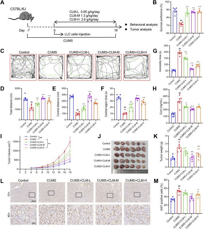
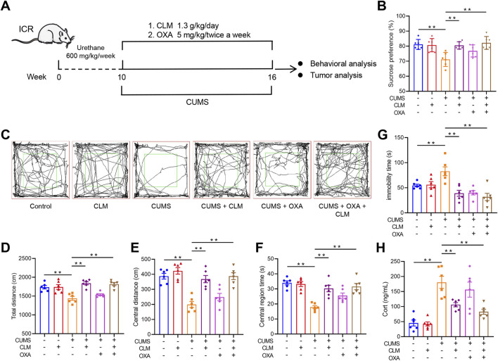
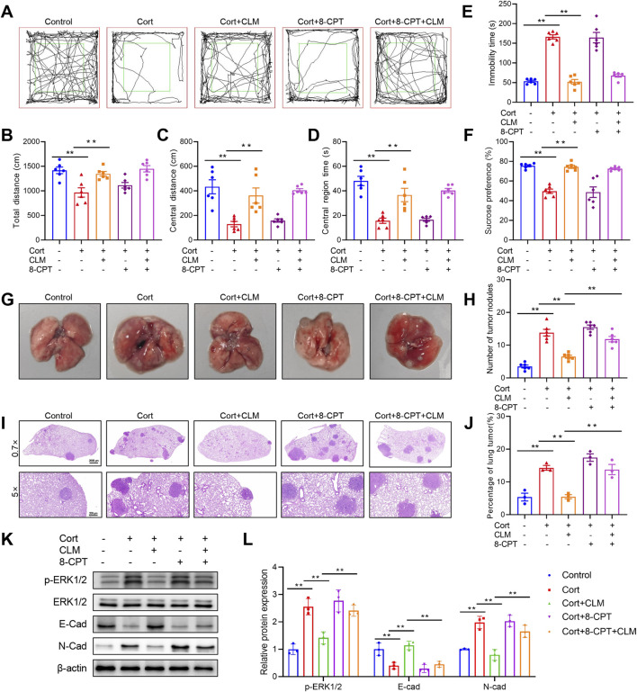
Methods: A lung cancer cell xenograft model combined with chronic unpredictable mild stress (CUMS) was used to evaluate the effect of CLM on lung cancer growth. Proteomic analysis was performed to explore the underlying mechanisms by which CLM alleviates CUMS-induced lung cancer progression. Western blot and qPCR were conducted to detect changes in Rap1/ERK-mediated epithelial-mesenchymal transition (EMT) progression. Finally, Rap1 agonists were utilized to determine the therapeutic mechanism of CLM on cortisol or corticosterone (Cort)-induced EMT progression in lung cancer cells and a mouse lung cancer model.

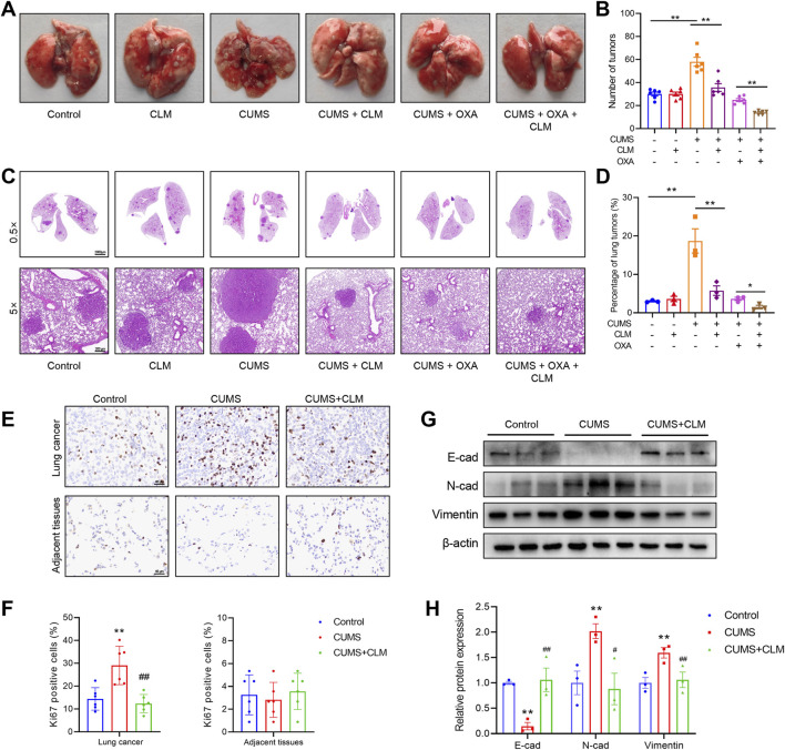
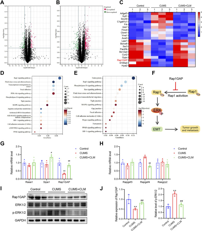
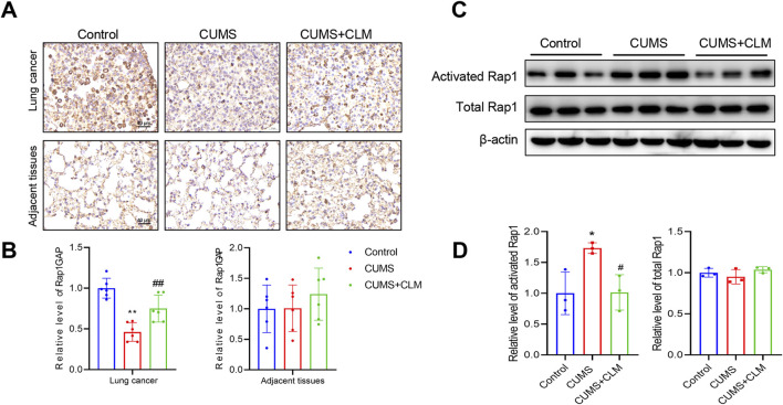
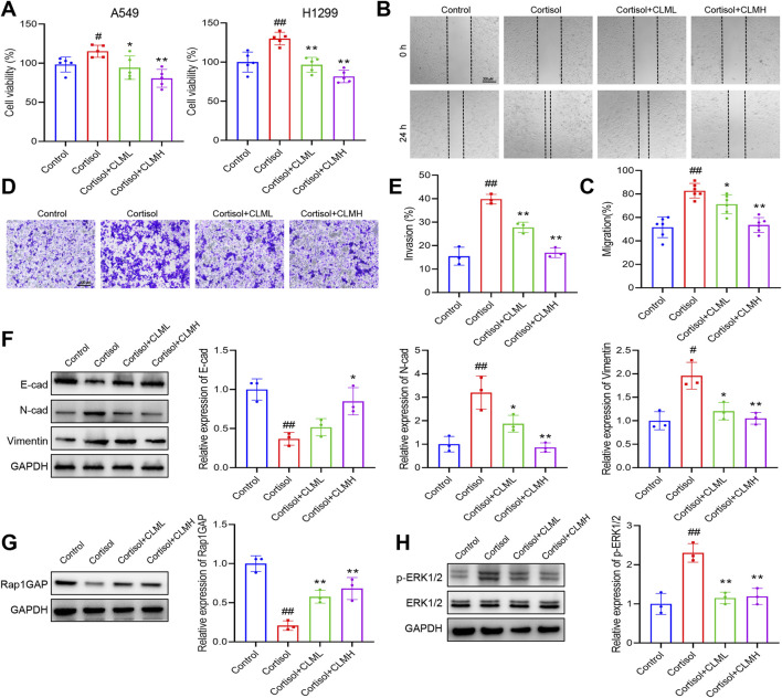
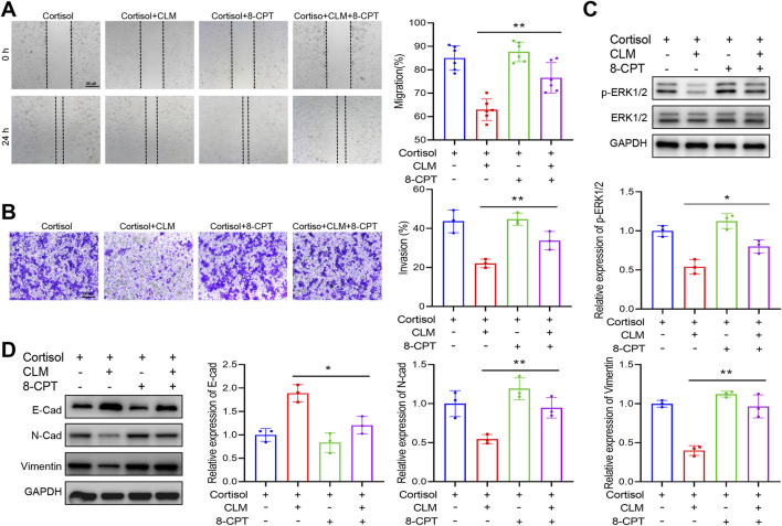
Results: In our study, CUMS promoted lung cancer xenograft growth, increased the expression of the proliferation marker Ki67, and elevated serum Cort levels. CLM treatment not only alleviated CUMS-induced depression-like behaviors, but also suppressed stress-driven tumor growth. These effects were replicated in a urethane-induced lung cancer model combined with CUMS. Proteomic analysis revealed that CLM's anti-tumor effects were associated with modulation of the Rap1 pathway. Mechanistically, CUMS downregulated Rap1GAP, activating Rap1 and subsequent ERK1/2 phosphorylation, thereby promoting EMT in lung cancer tissues. CLM effectively reversed these effects by inhibiting Rap1/ERK-mediated EMT. In vitro, CLM suppressed cortisol-induced migration, invasion, and EMT in lung cancer cells, and these effects were attenuated by Rap1 agonists. Furthermore, CLM inhibited Cort-induced EMT and depression-like behaviors in vivo, while Rap1 activation diminished CLM's efficacy against Cort-driven tumor growth.

Conclusion: These findings suggest that Rap1/ERK-mediated EMT is a hallmark of chronic stress-associated lung cancer progression. CLM exerts its therapeutic effects by targeting this pathway, offering a novel strategy to mitigate stress-aggravated oncogenesis.

Keywords: Chaihu Longgu Muli Decoction; Rap1; chronic stress; epithelial-mesenchymal transition; lung cancer.


Conflict of interest statement

The authors declare that the research was conducted in the absence of any commercial or financial relationships that could be construed as a potential conflict of interest.

Figures

FIGURE 1
FIGURE 1
Representative base peak chromatograms of CLM. (A) Profile acquired in positive ionization mode. (B) Profile acquired in negative ionization mode.
FIGURE 2
FIGURE 2
CLM improves depression like behavior and inhibits transplantation tumor growth induced by CUMS in mice. (A) Schematic of CUMS exposure of the lung tumor model. (B) Sucrose preference of tumor bearing mice (n = 6). (C) Trajectory diagram of open field test. (D) The total distance of mice in open field (n = 6). (E) The central distance of mice in open field (n = 6). (F) The central region time of mice in open field (n = 6). (G) The immobility time of tail suspension test (n = 6). (H) The serum Cort content of mice was detected by the ELISA kit (n = 6). (I) Tumor growth curve of tumor bearing mice in different groups. (J) Photos of transplanted tumors (n = 6). (K) Tumor weight (n = 6). (L) Representative photos of Ki67 immunohistochemical staining. (M) Statistical analysis of Ki67 positive cell rate (n = 6). Data are represented as mean ± SEM. *P < 0.05, **P < 0.01, compared with CUMS group; ## P < 0.01, compared with control group.
FIGURE 3
FIGURE 3
CLM improves depression like behavior in urethane induced lung cancer combined with CUMS in mice. (A) Schematic of CUMS exposure of the lung cancer model. (B) Sucrose preference of tumor bearing mice (n = 6). (C) Trajectory diagram of open field test (n = 6). (D) The total distance of mice in open field (n = 6). (E) The central distance of mice in open field (n = 6). (F) The central region time of mice in open field (n = 6). (G) The immobility time of tail suspension test (n = 6). (H) The serum Cort content of mice was detected by the ELISA kit (n = 6). Data are represented as mean ± SEM. **P < 0.01.
FIGURE 4
FIGURE 4
CLM inhibits lung cancer growth induced by CUMS in mice. (A) Representative photos of mouse lung tissues. (B) Statistical analysis of lung tumor number in mice (n = 6). (C) Representative pictures of HE staining of mouse lung tissues. (D) Statistical analysis of tumor area in mouse lung tissues (n = 3). (E) Representative photos of Ki67 immunohistochemical staining (40×). (F) Statistical analysis of Ki67 positive cell rate (n = 6). (G) Western blotting analysis of the expression of E-cad, N-cad, and Vimentin in lung tissues (n = 3). (H) Statistical analysis of Western blot (n = 3). Data are represented as mean ± SEM. *P < 0.05, **P < 0.01, compared with control group; ## P < 0.01, compared with CUMS group.
FIGURE 5
FIGURE 5
CLM inhibits the activation of Rap1/ERK pathway induced by CUMS. (A) The volcanic plots represent the relative expressions of differentially expressed proteins, CUMS group v. s. Control group. (B) The volcanic plots represent the relative expressions of differentially expressed proteins, CUMS + CLM group v. s. CUMS group. (C) The heat map of top 20 differentially expressed proteins. (D) The KEGG pathway enrichment results of differentially expressed proteins in CUMS and control group. (E) The KEGG pathway enrichment results of differentially expressed proteins in CUMS + CLM and CUMS group. (F) Schematic diagram of signal pathway transduction. (G) The expression of Rap1 repressed related genes was detected by qPCR (n = 3). (H) The expression of Rap1 activation related genes was detected by qPCR (n = 3). (I) Western blotting analysis of the expression of Rap1GAP, ERK, and p-ERK in lung tissues. (J) Statistical analysis of Western blot (n = 3). Data are represented as mean ± SEM. *P < 0.05, **P < 0.01, compared with control group; ## P < 0.01, compared with CUMS group.
FIGURE 6
FIGURE 6
CLM inhibit the activation of Rap1 induced by CUMS. (A) Representative photos of Rap1GAP immunohistochemical staining (40×). (B) Statistical analysis of Rap1GAP level (n = 6). (C) The levels of activated Rap1 and total Rap1 were detected by Western blot (n = 3). (D) Statistical analysis of Western blot (n = 3). Data are represented as mean ± SEM. *P < 0.05, **P < 0.01, compared with control group; # P < 0.05, ## P < 0.01, compared with CUMS group.
FIGURE 7
FIGURE 7
CLM inhibits cortisol induced EMT progression of lung cancer cells. (A) The cell viability analysis of A549 or H1299 cells treated with cortisol (2 μM), cortisol (2 μM) + CLML (50 μg/mL), and Cort (2 μM)+CLMH (100 μg/mL) for 48 h (n = 5). (B,C) The cell migration ability was assessed using the scratch assay (n = 6). (D,E) The cell invasion ability was assessed using the transwell assay (n = 3). (F) The protein levels of E-cad, N-cad, and Vimentin in H1299 cells was measured by Western blot (n = 3). (G,H) The protein levels of Rap1GAP, ERK1/2, and p-ERK1/2 in H1299 cells was measured by Western blot (n = 3). Data are represented as mean ± SEM. *P < 0.05, **P < 0.01, compared with cortisol group; # P < 0.05, ## P < 0.01, compared with control group.
FIGURE 8
FIGURE 8
Rap1 activator reverses the effect of CLM on cortisol induced migration, invasion. (A) The cell migration ability was assessed using the scratch assay (n = 6). (B) The cell invasion ability was assessed using the transwell assay (n = 3). (C) The protein levels of ERK1/2, and p-ERK1/2 in H1299 cells was measured by Western blot (n = 3). (D) The protein levels of E-cad, N-cad, and Vimentin in H1299 cells was measured by Western blot (n = 3). Data are represented as mean ± SEM. *P < 0.05, **P < 0.01.
FIGURE 9
FIGURE 9
Rap1 activator reverses the efficacy of CLM in the inhibiton of Cort induced lung cancer progress. (A) Trajectory diagram of open field test. (B) The total distance of mice in open field (n = 6). (C) The central distance of mice in open field (n = 6). (D) The central region time of mice in open field (n = 6). (E) The immobility time of tail suspension test (n = 6). (F) Sucrose preference of tumor bearing mice (n = 6). (G) Representative photos of mouse lung tissues. (H) Statistical analysis of lung tumor number in mice (n = 6). (I) Representative pictures of HE staining of mouse lung tissues. (J) Statistical analysis of tumor area in mouse lung tissues (n = 3). (K,L) Western blotting analysis of the expression of E-cad, N-cad, ERK1/2, and p-ERK1/2 in lung tissues (n = 3). Data are represented as mean ± SEM. **P < 0.01.

Similar articles

References

    1. An Y., Liu Z., Wang S., Wang Q., Zhang C., Zhang L., et al. (2022). Effect of chaihu plus longgu muli decoction plus five-element music therapy in the treatment of cancer-related depression. Support. Care Cancer Off. J. Multinatl. Assoc. Support. Care Cancer 30, 7955–7962. 10.1007/s00520-022-07172-6 - DOI - PubMed
    1. Bortolato B., Hyphantis T. N., Valpione S., Perini G., Maes M., Morris G., et al. (2017). Depression in cancer: the many biobehavioral pathways driving tumor progression. Cancer Treat. Rev. 52, 58–70. 10.1016/j.ctrv.2016.11.004 - DOI - PubMed
    1. Cui B., Peng F., Lu J., He B., Su Q., Luo H., et al. (2021a). Cancer and stress: NextGen strategies. Brain. Behav. Immun. 93, 368–383. 10.1016/j.bbi.2020.11.005 - DOI - PubMed
    1. Cui S., Lin H., Cui Y., Wen W., Cui X., Shen C., et al. (2021b). Depression promotes lung carcinoma progression by regulating the tumor microenvironment in tumor-bearing models of C57BL/6J mice. Neurosci. Lett. 754, 135851. 10.1016/j.neulet.2021.135851 - DOI - PubMed
    1. Dai S., Mo Y., Wang Y., Xiang B., Liao Q., Zhou M., et al. (2020). Chronic stress promotes cancer development. Front. Oncol. 10, 1492. 10.3389/fonc.2020.01492 - DOI - PMC - PubMed

LinkOut - more resources