Review
. 2025 Jul 25:16:1609033.
doi: 10.3389/fneur.2025.1609033. eCollection 2025.

Technological advances in the diagnosis and management of inherited optic neuropathies

Affiliations
Review

Technological advances in the diagnosis and management of inherited optic neuropathies

John O T Britton et al. Front Neurol. .

Abstract

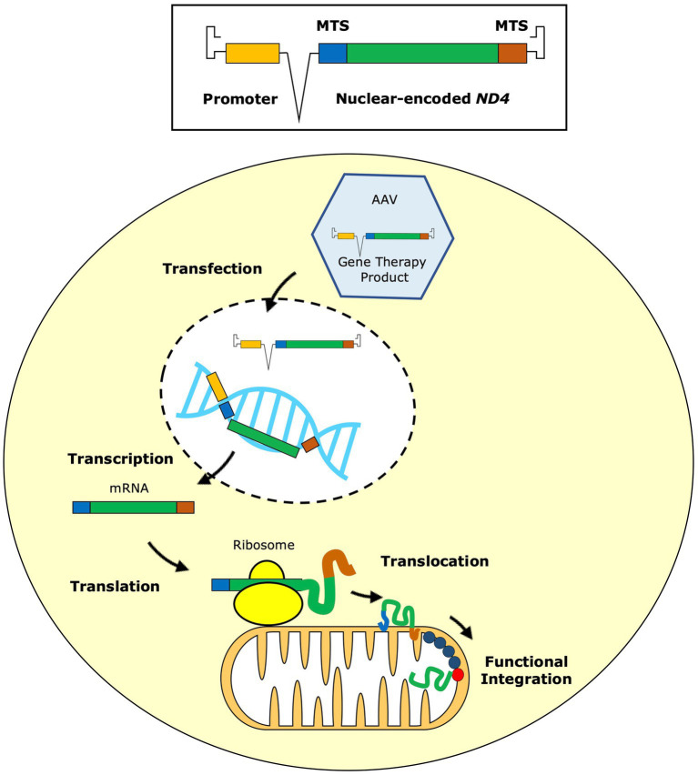
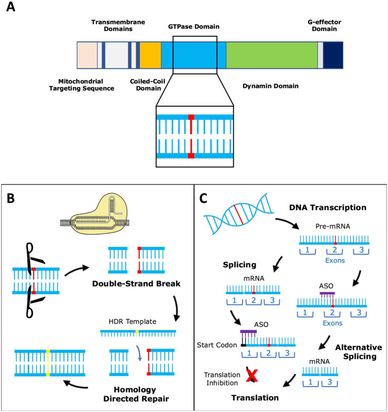
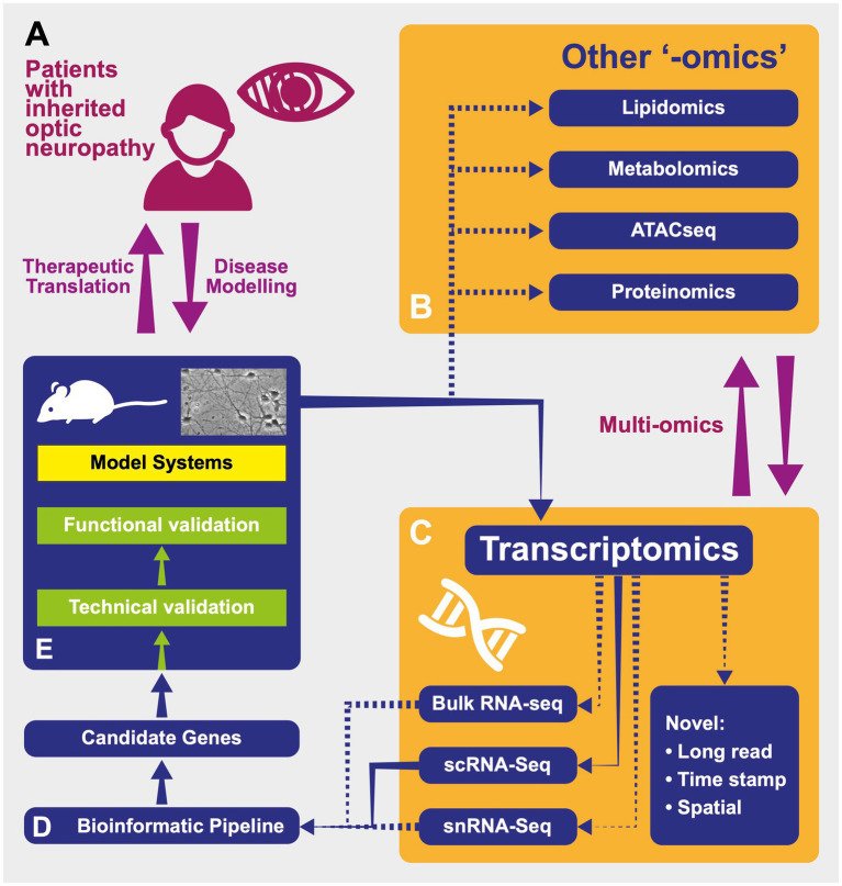
Preferential degeneration of retinal ganglion cells (RGCs) is a defining feature of the inherited optic neuropathies (IONs), a group of monogenic eye diseases predominately comprising Leber hereditary optic neuropathy (LHON) and autosomal dominant optic atrophy (DOA). Their pathogenesis is characterised by mitochondrial dysfunction, which causes loss of RGCs leading to irreversible vision loss. Although currently incurable, there are several emerging therapeutic avenues encompassing gene therapies, precision medicine strategies and neuroprotection. These are underscored by recent technological advances such as next-generation sequencing and improved disease modelling. In this review, we discuss these advances and the impact these will have on future diagnostic and treatment capabilities. We first focus on the clinical presentation and pathogenic mechanisms of LHON and DOA, followed by a discussion of emerging technology to facilitate diagnosis and treatment. We highlight the current unmet clinical demand of IONs, and the promise of current and future research developments.

Keywords: autosomal dominant; hereditary; leber; optic atrophy; retinal ganglion cells.


Conflict of interest statement

The authors declare that the research was conducted in the absence of any commercial or financial relationships that could be construed as a potential conflict of interest.

Figures

Figure 1
Figure 1
Reproduced with permission from Chen et al. (2023). The gene therapy vector contains the replacement mitochondrial ND4 gene modified according to the nuclear code. This nuclear-encoded version of ND4 is fused to two mitochondrial targeting sequences (MTS) and a promoter is inserted for more efficient expression. The gene therapy vector is packaged into an adeno-associated virus (AAV), usually serotype 2, which is delivered to the retinal ganglion cells by intravitreal injection. The AAV transfects the retinal ganglion cells, delivering the gene therapy product into the nucleus, where it remains in an episomal state. From here, the inserted gene sequence undergoes transcription to messenger RNA (mRNA) and, with the aid of the MTS, it is directed to mitochondrial ribosomes on the external surface of the mitochondrial outer membrane, where gene translation occurs. As it is being synthesised, the newly translated ND4 protein is translocated across the two mitochondrial membranes into the mitochondrial matrix aided by the MTS. Finally, the imported ND4 subunit undergoes post-translational folding before being integrated within complex I, restoring its function.
Figure 2
Figure 2
Reproduced with permission from Chen et al. (2023). (A) The OPA1 gene codes for a large multimeric GTPase that localises to the mitochondrial inner membrane and regulates a number of important mitochondrial functions. Over 500 pathogenic variants (represented by the red base pair) of OPA1 have been reported and these span the entire length of the gene. (B) OPA1 variants can be corrected by gene editing using CRISPR-Cas9. Double-strand breaks in DNA are introduced by Cas9 and the correct base sequence is inserted by homology directed repair (HDR) using an HDR template. (C) Another strategy is to modify OPA1 expression by altering gene transcription using antisense oligonucleotides (ASOs). ASOs can bind to mRNA near the translation start site and either enhance or suppress translation. Alternatively, the ASO can bind to pre-mRNA resulting in alternative splicing, which could be used to restore normal splicing in the setting of OPA1 splice variants.
Figure 3
Figure 3
Reproduced with permission from Gilhooley et al. (74). A schematic representing the processes of modelling and investigating inherited optic neuropathies using “-omics” methods. (A) Model diseases such as DOA and LHON can be powerful tools in which to investigate the effects of mitochondrial dysfunction. The study of patients with inherited optic neuropathies is often a two-way process: in one direction, the characterisation of their phenotype and genotype allows the development of useful disease models (for example, cell- and animal-based). These provide an efficient environment in which to increase our understanding of underlying disease processes as well as a testing ground for novel therapies before their return in the opposite direction, back into patients. (B) Tissues from model systems can be investigated using multiple “-omics” techniques—in some cases, these can be performed simultaneously and synthesised into a multiomic approach. ATACseq—Assay for Transposase Accessible Chromatin—a method for assessing which areas of the chromatin superstructure are open and so likely available for active in transcription, is an example of the rapidly expanding field of epigenomics. (C) Multiple transcriptomic techniques are available and have particular strength in different experimental situations. “Novel”: Novel techniques are emerging that will further the resolution of these techniques including those with abilities to sequence longer transcripts in one read and those that integrate temporal and spatial information regarding transcripts (see text). RNA-seq—RNA-sequencing; scRNA-seq—single-cell RNAseq; snRNAseq—single nucleus RNAseq. (D) Transcriptomic techniques gain power from the large quantity of data that they produce. This necessitates adequately designed bioinformatic pipelines that are tailored to the exact scientific question being asked in order to produce a list of candidate genes for further investigation back in model systems. (E) Model systems of diseases can include those based on cultured patient cells or be animals carrying pathogenic variants, leading to phenocopies of human disease. Samples from these models used in transcriptomic analysis can include tissue (such as retinas) or cell cultures.

Similar articles

References

    1. Barboni P, Savini G, Parisi V, Carbonelli M, La Morgia C, Maresca A, et al. Retinal nerve fiber layer thickness in dominant optic atrophy: measurements by optical coherence tomography and correlation with age. Ophthalmology. (2011) 118:2076–80. doi: 10.1016/j.ophtha.2011.02.027 - DOI - PubMed
    1. Spiegel SJ, Sadun AA. Solutions to a radical problem: overview of current and future treatment strategies in Leber’s hereditary opic neuropathy. Int J Mol Sci. (2022) 23:13205. doi: 10.3390/ijms232113205, PMID: - DOI - PMC - PubMed
    1. Ito YA, Di Polo A. Mitochondrial dynamics, transport, and quality control: a bottleneck for retinal ganglion cell viability in optic neuropathies. Mitochondrion. (2017) 36:186–92. doi: 10.1016/j.mito.2017.08.014, PMID: - DOI - PubMed
    1. Kim US, Mahroo OA, Mollon JD, Yu-Wai-Man P. Retinal ganglion cells—diversity of cell types and clinical relevance. Front Neurol. (2021) 12:661938. doi: 10.3389/fneur.2021.661938, PMID: - DOI - PMC - PubMed
    1. Nissen C, Rönnbäck C, Sander B, Herbst K, Milea D, Larsen M, et al. Dissociation of pupillary post-illumination responses from visual function in confirmed OPA1 c.983A > G and c.2708_2711delTTAG autosomal dominant optic atrophy. Front Neurol. (2015) 6:5. doi: 10.3389/fneur.2015.00005 - DOI - PMC - PubMed

LinkOut - more resources