Wake-up intracerebral hemorrhage: hematoma expansion and outcomes
- PMID: 40786634
- PMCID: PMC12331591
- DOI: 10.3389/fneur.2025.1620170
Wake-up intracerebral hemorrhage: hematoma expansion and outcomes
Abstract
Introduction: While understudied, wake-up intracerebral hemorrhage (WU-ICH) is not uncommon (8.8-20.3% of ICH patients). Since the risk of hematoma expansion (HE) decreases as time passes, an uncertain onset time in WU-ICH may influence the risk of in-hospital HE and the potential effects of HE-preventive treatments. We aimed to evaluate HE and outcomes in WU-ICH compared to known-onset ICH.
Methods: We included ICH patients admitted to the Karolinska University Hospital from 2016 to 2022, comparing WU-ICH vs. known-onset ICH regarding baseline characteristics, HE, and outcomes.
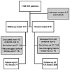
Results: Of 763 patients, 147 (19%) had WU-ICH and 616 (81%) had known onset, median (IQR) last-known-well to hospital time 9.6 h (5.9-12.2 h) vs. 1.3 h (0.9-2.0 h). WU-ICH patients more often had dementia (15% vs. 5%, p < 0.001), oral anticoagulants (26% vs. 16%, p = 0.005), and pre-stroke modified Rankin Scale 3-5 (24% vs. 15%, p = 0.01). Baseline ICH volume was 14 mL (6-35 mL) vs. 13 mL (5-34 mL). Among patients who underwent CT angiography at admission, 15% of WU-ICH vs. 27% of known-onset ICH had spot signs (p = 0.002). Of patients with CT follow-up <72 h, HE occurred in 24/77 (31.2%) in WU-ICH, and 123/356 (34.6%) in known-onset ICH, p = 0.57. Wake-up onset was not associated with HE in multivariable analysis, adjusted OR = 0.79 (95% CI 0.43-1.42). Analysis of the 3-month modified Rankin Scale showed no differences (median 4 vs. 4), unadjusted p = 0.35 and adjusted p = 0.78.
Conclusion: WU-ICH had a similar risk of HE and similar 3-month outcomes as known-onset ICH. Excluding WU-ICH from future trials targeting HE may be unwarranted.
Keywords: acute stroke; computed tomography; intracerebral hemorrhage; mortality; outcomes assessment.
Copyright © 2025 Almqvist, Delgado, Sjöstrand and Mazya.
Conflict of interest statement
The authors declare that the research was conducted in the absence of any commercial or financial relationships that could be construed as a potential conflict of interest.
Figures


Similar articles
-
Ultra-Early Hematoma Expansion Is Associated With Ongoing Hematoma Growth and Poor Functional Outcome.Stroke. 2025 Apr;56(4):838-847. doi: 10.1161/STROKEAHA.124.050131. Epub 2025 Jan 30. Stroke. 2025. PMID: 39882606 Clinical Trial.
-
Optimal Magnitude of Blood Pressure Reduction and Hematoma Growth and Functional Outcomes in Intracerebral Hemorrhage.Neurology. 2025 Mar 11;104(5):e213412. doi: 10.1212/WNL.0000000000213412. Epub 2025 Feb 6. Neurology. 2025. PMID: 39913881 Clinical Trial.
-
Influence of Hospital Type on Outcomes of Patients With Acute Spontaneous Intracerebral Hemorrhage: A Population-Based Study.Neurology. 2024 Jul 23;103(2):e209539. doi: 10.1212/WNL.0000000000209539. Epub 2024 Jun 14. Neurology. 2024. PMID: 38875516
-
Haemostatic therapies for stroke due to acute, spontaneous intracerebral haemorrhage.Cochrane Database Syst Rev. 2023 Oct 23;10(10):CD005951. doi: 10.1002/14651858.CD005951.pub5. Cochrane Database Syst Rev. 2023. PMID: 37870112 Free PMC article.
-
Antithrombotic treatment after stroke due to intracerebral haemorrhage.Cochrane Database Syst Rev. 2023 Jan 26;1(1):CD012144. doi: 10.1002/14651858.CD012144.pub3. Cochrane Database Syst Rev. 2023. PMID: 36700520 Free PMC article.
References
-
- Greenberg SM, Ziai WC, Cordonnier C, Dowlatshahi D, Francis B, Goldstein JN, et al. 2022 guideline for the Management of Patients with Spontaneous Intracerebral Hemorrhage: a guideline from the American Heart Association/American Stroke Association. Stroke. (2022) 53:e282–361. doi: 10.1161/STR.0000000000000407, PMID: - DOI - PubMed
LinkOut - more resources
Full Text Sources

