Case Reports
. 2025 Aug;15(8):140-144.
doi: 10.13107/jocr.2025.v15.i08.5916.

Vulvar Pressure Ulcer - An Inadvertent Complication of Thomas Splint: Case Report

Affiliations
Case Reports

Vulvar Pressure Ulcer - An Inadvertent Complication of Thomas Splint: Case Report

Alok Chandra Agrawal et al. J Orthop Case Rep. 2025 Aug.

Abstract

Introduction: Pressure ulcers are common complications in immobilized patients, typically occurring over bony prominences. However, in rare instances, particularly in high-risk individuals with hematologic disorders such as sickle cell disease, ulcers may develop at unusual anatomical sites due to external medical devices. This case highlights a rare presentation of a vulvar pressure ulcer caused by prolonged application of a Thomas splint in a patient with homozygous sickle cell disease (HbSS).

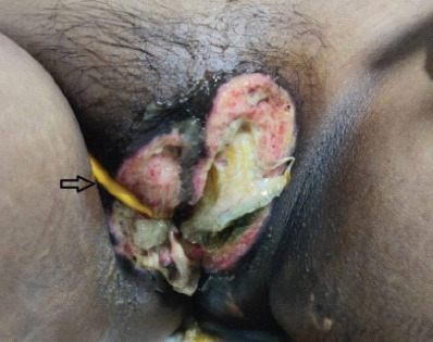
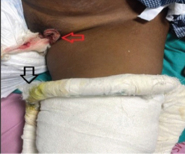
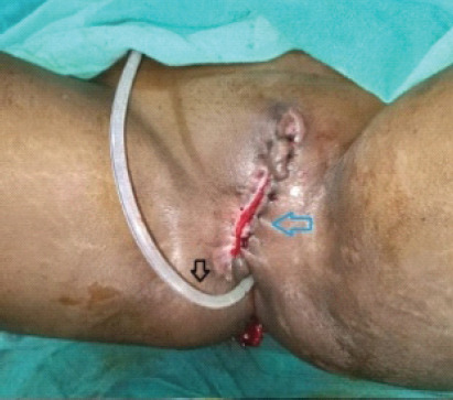
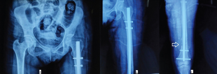
Case report: A 24-year-old woman with HbSS was admitted with a closed distal femoral shaft fracture. She was immobilized using a Thomas splint. On the 15th day of hospitalization, she developed a necrotic ulcer over the labia majora, diagnosed as a vulvar pressure ulcer secondary to device compression. Conservative wound management was initiated, followed by surgical debridement and closure using local advancement flaps. Orthopedic care was modified, and internal fixation of the fracture was later performed in a pressure-avoiding position. The wound healed completely within 2 weeks of surgery.

Conclusion: Vulvar pressure ulcers are rare and often underdiagnosed, particularly in patients with risk factors like sickle cell disease. This case emphasizes the need for regular inspection of pressure points, patient education, and multidisciplinary collaboration to prevent and manage such complications. Thoughtful application and monitoring of immobilization devices can reduce the risk of pressure injuries in vulnerable populations.

Keywords: Pressure ulcer; Thomas splint; orthopedic complication; sickle cell disease; vulvar wound.


Conflict of interest statement

Conflict of Interest: Nil

Figures

Figure 1
Figure 1
Pre-operative radiograph of right knee, including leg and thigh (lateral view), demonstrating a distal femoral shaft fracture. Legend: Black arrow indicates an oblique fracture in the distal shaft of the femur with minimal angulation and slight displacement.
Figure 2
Figure 2
Preoperative clinical photograph demonstrating a severe vulvar pressure ulcer involving the labia majora. The ulcer is characterized by extensive tissue necrosis, with visible yellowish slough and purulent discharge occupying the wound bed. Surrounding skin shows signs of inflammation and maceration, suggestive of ongoing infection and compromised local vascularity. Legend: The black arrow indicating indwelling foley’s catheter in situ.
Figure 3
Figure 3
Preoperative clinical photograph demonstrating the application of a Thomas splint with evident compression over the proximal thigh and groin region. Legend: The black arrow indicates discoloration and contamination of the splint padding with urine and feces. The red arrow highlights a developing pressure ulcer over the labia majora, likely secondary to prolonged compression and soiling in the region.
Figure 4
Figure 4
Clinical image demonstrating the medial rim of a Thomas splint, which shows marked yellowish-brown discoloration of the padding. This staining is likely due to prolonged contamination with urine and stool, reflecting poor perineal hygiene and inadequate splint care. Legend: The black arrow indicates the yellowish brown discolouration of Thomas splint padding.
Figure 5
Figure 5
Pre-operative clinical photograph of a vulval pressure ulcer following three sessions of surgical debridement. The wound bed appears clean, with no visible necrotic tissue or slough, and shows early signs of epithelialization along the wound margins. Legend: Black arrow indicates epithelializing wound edges; red arrow points to the indwelling silicone catheter in situ.
Figure 6
Figure 6
Post-operative clinical photograph demonstrating successful closure of the vulval pressure sore using local flap advancement. The flap was sutured without tension, ensuring proper wound edge approximation. Legend: Blue arrow indicates the flap suture line; black arrow points to the silicone catheter in situ.
Figure 7
Figure 7
Post-operative radiographs showing intramedullary interlocking nailing for distal femur fracture, demonstrating good fracture reduction, alignment, and stable fixation. (a) Pelvis with both hips in anteroposterior (AP) view reveals well-aligned hip joints, an intact pubic symphysis, and the femoral nail in situ on the left side. Proximal locking screws are appropriately positioned and of correct length. (b) AP radiograph of the left hip and thigh showing accurate placement of the femoral nail with maintained alignment and no signs of malrotation or displacement. (c) AP radiograph of the left distal femur and knee demonstrating proper reduction of the distal femur fracture, stabilized with distal locking screws of adequate length and positioning. Legend: The white arrow in (c) indicates the reduced fracture site, secured with the femoral nail in situ.

Similar articles

  • Automated devices for identifying peripheral arterial disease in people with leg ulceration: an evidence synthesis and cost-effectiveness analysis.
    Boyers D, Cruickshank M, Aucott L, Kennedy C, Manson P, Bachoo P, Brazzelli M. Boyers D, et al. Health Technol Assess. 2024 Aug;28(37):1-158. doi: 10.3310/TWCG3912. Health Technol Assess. 2024. PMID: 39186036 Free PMC article.
  • Management of urinary stones by experts in stone disease (ESD 2025).
    Papatsoris A, Geavlete B, Radavoi GD, Alameedee M, Almusafer M, Ather MH, Budia A, Cumpanas AA, Kiremi MC, Dellis A, Elhowairis M, Galán-Llopis JA, Geavlete P, Guimerà Garcia J, Isern B, Jinga V, Lopez JM, Mainez JA, Mitsogiannis I, Mora Christian J, Moussa M, Multescu R, Oguz Acar Y, Petkova K, Piñero A, Popov E, Ramos Cebrian M, Rascu S, Siener R, Sountoulides P, Stamatelou K, Syed J, Trinchieri A. Papatsoris A, et al. Arch Ital Urol Androl. 2025 Jun 30;97(2):14085. doi: 10.4081/aiua.2025.14085. Epub 2025 Jun 30. Arch Ital Urol Androl. 2025. PMID: 40583613 Review.
  • Endovenous ablation for venous leg ulcers.
    Cai PL, Hitchman LH, Mohamed AH, Smith GE, Chetter I, Carradice D. Cai PL, et al. Cochrane Database Syst Rev. 2023 Jul 27;7(7):CD009494. doi: 10.1002/14651858.CD009494.pub3. Cochrane Database Syst Rev. 2023. PMID: 37497816 Free PMC article.
  • Dressings and topical agents for treating venous leg ulcers.
    Norman G, Westby MJ, Rithalia AD, Stubbs N, Soares MO, Dumville JC. Norman G, et al. Cochrane Database Syst Rev. 2018 Jun 15;6(6):CD012583. doi: 10.1002/14651858.CD012583.pub2. Cochrane Database Syst Rev. 2018. PMID: 29906322 Free PMC article.
  • Compression for venous leg ulcers.
    O'Meara S, Cullum N, Nelson EA, Dumville JC. O'Meara S, et al. Cochrane Database Syst Rev. 2012 Nov 14;11(11):CD000265. doi: 10.1002/14651858.CD000265.pub3. Cochrane Database Syst Rev. 2012. PMID: 23152202 Free PMC article.

References

    1. Bergstrom N, Horn SD, Smout RJ, Bender SA, Ferguson ML, Taler G, et al. The national pressure ulcer long-term care study:Outcomes of pressure ulcer treatments in long-term care. J Am Geriatr Soc. 2005;53:1721–9. - PubMed
    1. Mancoll JS, Phillips LG. Pressure sores. In: Achauer BM, Erikson E, Guzuron B, Coleman J 3rd, Russell R, Vander Kolk C, editors. Plastic Surgery. Vol. 1. St. Louis, MO: Mosby Inc; 2000. pp. 447–62.
    1. Conde Montero E, Martínez Gómiz JM, De La Cueva Dobao P. The vulva:An uncommon presentation of a pressure ulcer. Wounds. 2017;29:E28–31. - PubMed
    1. Rakic VS, Colic MM, Lazovic GD. Unusual localisation of pressure ulcer--the vulva. Int Wound J. 2011;8:313–6. - PMC - PubMed
    1. Rathore FA, New PW, Waheed A. Pressure ulcers in spinal cord injury:An unusual site and etiology. Am J Phys Med Rehabil. 2009;88:587–90. - PubMed

Publication types

LinkOut - more resources