. 2025 Feb 7;9(4):407-415.
doi: 10.22603/ssrr.2024-0264. eCollection 2025 Jul 27.

Clinical and Imaging Characteristics of Patients with Cervical Compressive Myelopathy Presenting with Unilateral Motor Deficits

Affiliations

Clinical and Imaging Characteristics of Patients with Cervical Compressive Myelopathy Presenting with Unilateral Motor Deficits

Masatsune Sato et al. Spine Surg Relat Res. .

Abstract

Introduction: Cervical compressive myelopathy is a leading cause of spinal cord dysfunction in middle-aged and older adults. Although the pathological classification of cervical myelopathy is well established, the quantitative analysis of its imaging features remains underexplored. This study quantitatively evaluated the imaging characteristics of unilateral motor deficit cervical compressive myelopathy.

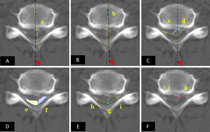
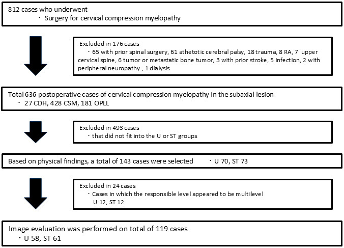
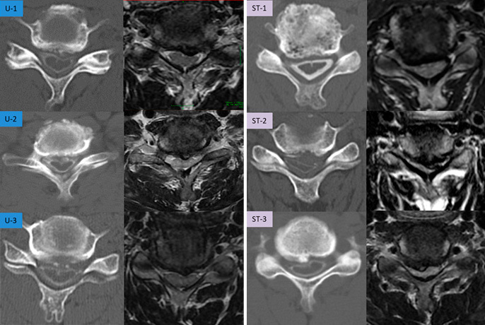
Methods: This retrospective observational study included patients who underwent surgery for cervical compressive myelopathy between 2009 and 2023. Pre-operative cervical magnetic resonance imaging (MRI) and postmyelographic computed tomography (CTM) axial images were assessed for spinal cord rotation, deformity, available space, and signal changes. Patients were classified into unilateral motor deficit (Group U) and symmetric transverse (Group ST) types, and were analyzed for specific imaging parameters.

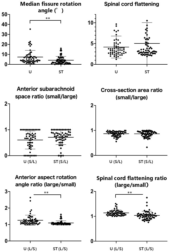
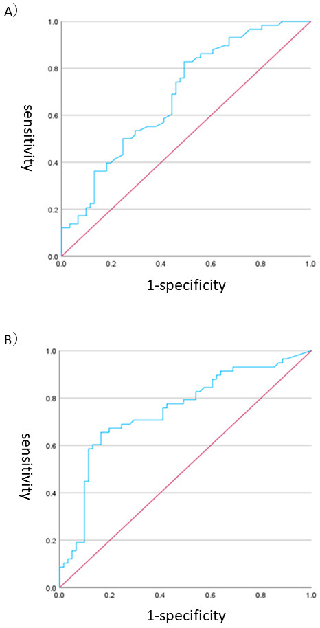
Results: The final analysis included 119 of the 812 identified patients. Group U patients were younger (59.1±13.8 years) and had higher Japanese Orthopaedic Association scores (10.6±2.7) compared with Group ST patients (71.1±11.0 years, 8.4±2.3). Group U showed significant morphological differences, including a reduced anterior-subarachnoid space and increased spinal cord rotation on the affected side. Group U exhibited significant differences in the median fissure rotation angle (7.4°±6.7°) and anterior-aspect rotation angle ratio (1.26±0.31) compared with Group ST (4.14°±3.87°, 1.10±0.14). Receiver operating characteristic curve analysis identified specific cutoff values for distinguishing Group U (2.80° for median fissure rotation angle and 1.116 for anterior-aspect rotation angle ratio). The MRI-based detection sensitivity was lower in Group U (27.6%) compared with in Group ST (68.9%).

Conclusions: Unilateral motor deficits are associated with distinctive spinal cord rotational deformities, including a greater median fissure rotation angle and anterior-aspect rotation angle ratio. CTM is better than MRI for detecting unilateral motor deficits. Future research to improve treatment outcomes should focus on spinal cord circulation assessment using advanced imaging techniques.

Keywords: cerebrospinal fluid space; cervical myelopathy; quantitative imaging analysis; spinal cord rotation; unilateral motor deficit.


Conflict of interest statement

Conflicts of Interest: The authors declare that there are no relevant conflicts of interest.

Figures

Figure 1.
Figure 1.
Imaging Analysis. A) Median fissure rotation angle (a°) at the affected level: the angle between the vertical line on the screen and the median fissure. B) Vertebral rotation angle (b°): the angle between the vertical line and the line connecting the vertebral body midline to the spinous process midline, measured for the vertebrae above and below the affected level (b°, b´°). C) Spinal cord anterior-aspect rotation angles for the left and right sides (c°, d°): the angle between the vertical line and the line connecting the median fissure to the inflection point of the anterior spinal cord curve. D) Spinal cord cross-sectional area e, f (mm2): cross-sectional area of the spinal cord divided by the median fissure. E) Longitudinal cord g (mm) and transverse diameters h and i (mm). F) Anterior-subarachnoid space j, k (mm). Based on these measurements, the corrected median fissure rotation angle=|a−(b+b´)/2| and the corrected spinal cord anterior-aspect rotation angle=|c−a|, |d−a|. Spinal cord flattening=(h+i)/g (whole), h/g (right), or i/g (left). The spinal cord anterior-aspect rotation angle ratio=larger side/smaller side. The cross-sectional area ratio=smaller side/larger side. The anterior-subarachnoid space ratio=smaller side/larger side. The flattening ratio=larger side/smaller side.
Figure 2.
Figure 2.
Patient Selection.
Figure 3.
Figure 3.
Intergroup Comparison of Imaging Measurements. Group U had significantly greater median fissure rotation angle (p<0.01), anterior-aspect rotation angle ratio (p<0.01), and spinal cord flattening ratio (p<0.01).
Figure 4.
Figure 4.
ROC Analysis for Group U. A) The receiver operating characteristic curve (ROC) using the median fissure rotation angle as an independent variable for determining group U showed a cutoff value of 2.80° based on Youden’s index (sensitivity 0.828; specificity 0.508). The area under the curve (AUC) was 0.684, with a 95% confidence interval of 0.589–0.78. The positive and negative predictive values were 61.2% and 66.7%, respectively. B) The ROC curve using the anterior-aspect rotation angle ratio as an independent variable for U group determination has a cutoff value of 1.116 (sensitivity 0.655; specificity 0.836). The AUC was 0.748, with a 95% confidence interval of 0.657–0.838. The positive and negative predictive values were 74.5% and 68.1%, respectively.
Figure 5.
Figure 5.
Case Presentations. The anterior-aspect rotation angle ratio was calculated as the larger side/smaller side; the anterior-subarachnoid space ratio was calculated as the smaller side/larger side. U-1 is a typical U group with increased median fissure and anterior-aspect rotation angles predominantly on the affected side. U-2 and U-3 exhibit motor impairment on the side opposite the predicted side, suggesting influences beyond static physical compression. ST-1 represents a typical ST-group case with a relatively small rotation angle. ST-2 shows a large median fissure rotation angle with marked bilateral atrophy, indicating spinal cord atrophy’s pathological role. ST-3 is clinically classified in Group ST; however, the myelographic computed tomography features resemble Group U despite magnetic resonance imaging showing bilateral signal changes. Age, sex (M: male, F: female), affected side (L: left, R: right; U group only), MRI signal change (L: left, R: right, B: bilateral, none), Japanese Orthopedic Association score, median fissure rotation angle (°), anterior-aspect rotation angle ratio, anterior-subarachnoid space ratio, spinal cord flattening ratio, and cross-sectional area (mm2) are U-1 (68, M, L, none, 9.5, 12.5°, 1.11, 0.66, 1.75, 31.3 mm2), U-2 (66, M, R, B, 10.5, 7.9°, 1.04, 0.25, 3.4, 42.5 mm2), U-3 (48, M, R, L, 10.5, 4°, 1.06, 1.0, 3.32, 50.7 mm2), ST-1 (71, F, -, B, 9.5, 3.3°, 1.08, 0.8, 2.76, 42 mm2), ST-2 (78, M, -, B, 7.5, 16.1°, 1.1, 0.8, 7.6, 20.9 mm2), and ST-3 (40, M, -, B, 9, 14.9°, 1.61, 0.75, 6.16, 50.7 mm2), respectively.

Similar articles

References

    1. Nurjck S. The pathogenesis of the spinal cord disorder associated with cervical spondylosis. Brain. 1972;95(1):87-100. - PubMed
    1. Vijay Kumar GR, Ray DK, Das RK. Natural history, prevalence, and pathophysiology of cervical spondylotic myelopathy. Indian Spine J. 2019;2(1):5-12.
    1. Development Committee for the Clinical Practice Guidelines on the Management of Cervical Spondylotic Myelopathy. The essence of clinical practice guidelines for cervical spondylotic myelopathy, 2020. Spine Surg Relat Res. 2024;8(2):119-32. - PMC - PubMed
    1. Breig A, Turnbull I, Hassler O. Effects of mechanical stresses on the spinal cord in cervical spondylosis. A study on fresh cadaver material. J Neurosurg. 1966;25(1):45-56. - PubMed
    1. White AA 3rd, Panjabi MM. Biomechanical considerations in the surgical management of cervical spondylotic myelopathy. Spine (Phila Pa 1976). 1988;13(7):856-60. - PubMed

LinkOut - more resources