Review
. 2025 Jul;86(4):483-500.
doi: 10.3348/jksr.2024.0107. Epub 2025 Jul 25.

Imaging Findings in Neurogenic Ptosis

Review

Imaging Findings in Neurogenic Ptosis

Yiseul Son et al. J Korean Soc Radiol. 2025 Jul.

Abstract

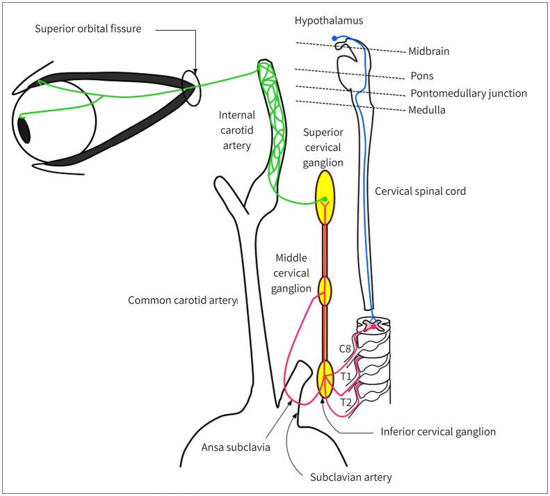
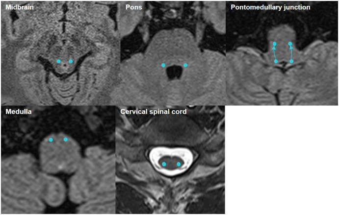
Neurogenic ptosis refers to upper eyelid drooping resulting from disrupted innervation of the eyelid retractor muscles. The differential diagnoses of neurogenic ptosis include oculomotor nerve palsy, Horner's syndrome, and neuromuscular junction disorders. Oculomotor nerve palsy and Horner's syndrome result from damage to the oculomotor and oculosympathetic pathways, respectively. The oculomotor nerve pathway has four segments: intraaxial, subarachnoid, cavernous, and orbital. The oculosympathetic pathway consists of three orders of neurons extending from the base of the skull to the upper chest. Several pathologic conditions can affect these neural pathways and cause neurogenic ptosis owing to the long course of the involved nerves. Therefore, neurogenic ptosis is usually associated with unique clinical features based on the anatomical location of the pathology. This pictorial essay provides a deeper understanding of the neural pathways and different diseases that cause neurogenic ptosis, which can help in determining the location of the pathology.

신경성 안검하수는 눈꺼풀을 들어 올리는 근육을 지배하는 신경에 문제가 생겨 눈꺼풀이 처지는 것을 말한다. 신경성 안검하수의 감별진단에는 안구운동신경마비, 호너증후군, 중증근육무력증을 포함한 신경근 접합부 장애 등이 있다. 특히 안구운동신경마비와 호너증후군은 각각 안구운동신경 경로와 안교감신경 경로의 손상에 의해 발생한다. 안구운동신경 경로는 축내분절, 지주막하분절, 해면분절, 안와분절의 4개 분절로 구성된다. 안교감신경 경로는 1차, 2차, 3차 뉴런으로 구성되며 두개골 기저부에서부터 가슴 상부에 걸쳐 있다. 이 신경들은 긴 경로로 이루어져 있기 때문에 다양한 질환들이 이 신경 경로에 영향을 미칠 수 있으며, 보통 해부학적 위치에 따라 독특한 임상적 특징들과 관련이 있다. 이 임상화보의 목적은 신경성 안검하수을 일으키는 신경 경로와 질병의 종류를 이해하고, 신경성 안검하수의 원인이 되는 위치를 유추할 수 있도록 하는 것이다.

Keywords: Anatomy; Horner’s Syndrome; Imaging; Oculomotor Nerve Palsy; Ptosis.


Conflict of interest statement

Conflicts of Interest: The authors have no potential conflicts of interest to disclose.

Figures

Fig. 1
Fig. 1. Upper eyelid anatomy.
The levator palpebrae superioris is the primary retractor muscle of the upper eyelid, which inserts onto the tarsal plate through the levator aponeurosis. Müller’s muscle originates from the levator palpebrae superioris, inserts onto the tarsal plate, and helps the levator palpebrae superioris to raise the upper eyelid.
Fig. 2
Fig. 2. Marginal reflex distance-1.
It is the distance between the margin of the upper eyelid and the central light reflex of the pupil. It is helpful for the diagnosis and evaluation of ptosis.
Fig. 3
Fig. 3. The oculomotor nerve pathway.
It consists of intra-axial, subarachnoid, cavernous, and orbital segments. The oculomotor nerve originates from the oculomotor nucleus located ventral to the cerebral aqueduct in the midbrain at the superior colliculus level. The subnucleus innervating the levator palpebrae superioris is a solitary nucleus located centrally and caudally. The other subnuclei are positioned bilaterally. The Edinger-Westphal nucleus—the accessory oculomotor nerve nucleus—is located in the rostral portion of the oculomotor nucleus. The oculomotor nerve passes between the PCA and SCA while exiting the midbrain, runs through the cavernous sinus, and enters the orbital fossa through the superior orbital fissure. PCA = posterior cerebral artery, SCA = superior cerebellar artery
Fig. 4
Fig. 4. Course of the intra-axial segment of the oculomotor nerve on axial T2-FLAIR image.
The nerve originates from the oculomotor nucleus in the midbrain (dots), and exits the brainstem ventrally passing through the red nucleus (lines).
Fig. 5
Fig. 5. Acute infarction in the midbrain.
A, B. A 48-year-old woman presented with left ptosis. Diffusion-weighted imaging (A) and apparent diffusion coefficient map (B) show a focal diffusion-restricted lesion in the left paramedian midbrain (arrows).
Fig. 6
Fig. 6. Course of the subarachnoid segment of the oculomotor nerve on axial T2 weighted VISTA image.
The nerve (arrows) emerges from the midbrain, medial to the cerebral peduncles. Thereafter, it runs forward and laterally between the posterior cerebral artery and superior cerebellar artery. VISTA = volume isotropic turbo spin-echo acquisition
Fig. 7
Fig. 7. SAH due to the rupture of a left posterior communicating artery aneurysm.
A, B. A 51-year-old woman presented with left third nerve palsy and ipsilateral ptosis. Brain CT angiography (A) shows a left posterior communicating artery aneurysm (arrow). SAH was not observed on CT (not shown), but cisternal and sulcal SAH is visible on susceptibility weighted imaging (B). SAH = subacute subarachnoid hemorrhage
Fig. 8
Fig. 8. Coronal contrast-enhanced T1-weighted VISTA image (A) shows the cavernous segment of the CN III (arrows). The sagittal T2 weighted image (B) shows the whole course of the CN III from the oculomotor nucleus to the levator palpebrae superioris-superior rectus complex.
CN III = oculomotor nerve, PCA = posterior cerebral artery, SCA = superior cerebellar artery, VISTA = volume isotropic turbo spin-echo acquisition
Fig. 9
Fig. 9. Traumatic carotid-cavernous fistula.
A, B. A 41-year-old man presented with right ptosis. Time-of-flight MR angiography (A) and its source image (B) show enlargement of both cavernous sinuses with the arterialized flow of high signal intensity (arrows) and marked dilatation of both superior ophthalmic veins (arrowheads).
Fig. 10
Fig. 10. Neurogenic tumor in the left cavernous sinus.
A, B. An 85-year-old woman presented with left ptosis and diplopia. Axial T1 (A) and T2 (B) weighted images show a well-defined T1 isointense and T2 inhomogeneously isointense nodular mass with a few T2 hyperintense foci in the left cavernous sinus (arrows). C. The mass demonstrates inhomogeneous mild enhancement on contrast-enhanced T1-weighted image (arrow).
Fig. 11
Fig. 11. Cavernous sinus and dural metastases from lung cancer.
A-C. A 72-year-old man presented with right ptosis. Contrast-enhanced axial (A, B) and coronal (C) T1-weighted images show enlargement of the right cavernous sinus with strong enhancement (arrows), which extends into the right orbital apex and sphenoid and ethmoid sinuses, along with diffuse uneven dural metastatic lesions in both cerebral convexities and the falx cerebri (empty arrows).
Fig. 12
Fig. 12. Orbital lymphoma.
A, B. A 58-year-old man presented with left ptosis. A well-defined homogeneous-enhancing soft tissue mass is seen in the left superior orbital fossa (arrows) with obliteration of the superior rectus muscle complex on coronal contrast-enhanced CT (A) and coronal contrast-enhanced T1-weighted image (B).
Fig. 13
Fig. 13. ANCA-associated vasculitis with central nervous system and renal involvement.
A, B. A 75-year-old woman presented with right ptosis. Axial T2-weighted (A) and contrast-enhanced T1-weighted (B) images show an expansile lesion with strong enhancement in the right cavernous sinus (arrows). The right distal internal carotid artery shows loss of normal vascular signal void with rim enhancement on axial contrast-enhanced T1-weighted image, indicating vasculitis. ANCA = anti-neutrophil cytoplasmic antibody
Fig. 14
Fig. 14. Invasive aspergillosis with cavernous sinus involvement.
A-C. An 80-year-old woman presented with mild left ptosis. CT (A) shows opacification in the left sphenoidal sinus with erosion of the adjacent bone (empty arrow). Axial T2 weighted (B) and axial contrast-enhanced T1-weighted (C) images show mucosal thickening with strong enhancement and central poorly-enhancing dark signal in the left sphenoidal sinus (arrows); the lesion infiltrates the left orbital apex, orbital fissures, and, cavernous sinus (arrowheads).
Fig. 15
Fig. 15. Pathway of the oculosympathetic nerve. The schematic diagram shows the course of the oculosympathetic nerve from the hypothalamus to the orbit. The first-order neurons (blue line) arise from the hypothalamus and descend through the brainstem and the spinal cord before synapsing in the ciliospinal center located at C8 to T2 levels. The second-order neurons (pink line) exit the spinal cord through the ventral roots and ascend through the inferior and middle cervical ganglia toward the superior cervical ganglion. The inferior and middle cervical ganglia are connected by two or more cords composed of anterior (also known as ansa subclavia) and posterior loops. The anterior loops run above the cervical pleura and loop beneath the first portion of the subclavian artery. The third-order neurons (green line) originate from the superior cervical ganglion and ascend along the internal carotid artery to form a nerve plexus. Finally, the nerve enters the orbital fossa through the superior orbital fissure, innervating the Müller’s and pupil dilator muscles.
Fig. 16
Fig. 16. Cross-sectional images of the oculosympathetic first-order neurons. The nerve fibers arising from the hypothalamus descend through the midbrain and pass lateral to the trochlear nucleus. At the pontomedullary junction, the fibers travel anteriorly from the area of the locus coeruleus to the ventral area of the ipsilateral inferior olivary nucleus. Then, the neurons descend lateral to the dorsal gray matter through the spinal cord to synapse in the ciliospinal center located at C8 to T2 levels.
Fig. 17
Fig. 17. Lateral medullary stroke.
A, B. A 58-year-old woman presented with right Horner’s syndrome, including right ptosis. Diffusion-weighted imaging (A) and apparent diffusion coefficient map (B) show a focal diffusion-restricted lesion in the right medulla (arrows), suggesting central Horner’s syndrome.
Fig. 18
Fig. 18. Images of the second-order oculosympathetic neuronal pathway.
A. T2-weighted image shows preganglionic neurons exiting through the C8 ventral spinal root. B-D. CT images show the ansa subclavia that passes above the cervical pleura and loops just below the first part of the subclavian artery (arrows). E. CT cross-sectional image shows the cervical sympathetic chain (dots) around the carotid sheath, anterior to the longus colli muscle (empty arrows).
Fig. 19
Fig. 19. Multiple metastatic neck lymph nodes.
A, B. A 53-year-old man with hepatocellular carcinoma and multiple neck lymph nodes metastases presented with left Horner’s syndrome, including left ptosis. Whole body PET-CT (A) and neck CT (B) show lymph node metastases (arrows) in the left supraclavicular area, presumably being the cause of left preganglionic Horner’s syndrome.
Fig. 20
Fig. 20. Iatrogenic trauma caused by surgical procedure.
A, B. A 60-year-old woman presented with left ptosis that developed after partial arch replacement surgery for aortic dissection (A). Postoperative CT (B) shows a hematoma around the left subclavian artery (arrow), suggesting preganglionic Horner’s syndrome.
Fig. 21
Fig. 21. Cross-sectional images of the oculosympathetic third-order neurons. The third-order neurons ascend along the internal carotid artery, forming a nerve plexus (dots). Subsequently, they pass through the cavernous sinus and enter the orbit through the superior orbital fissure (arrows).
Fig. 22
Fig. 22. ICA dissection.
A, B. A 59-year-old man with left ICA dissection presented with left Horner’s syndrome, including left ptosis. On coronal T1-weighted image (A) and source image of TOF MRA (B), left ICA shows double lumen with an intramural hematoma (arrows). C. TOF MRA shows mild luminal narrowing of the left ICA (arrow), suggesting postganglionic Horner’s syndrome. ICA = internal carotid artery, MRA = MR angiography, TOF = time-of-flight

Similar articles

  • Ptosis Correction.
    Koka K, Patel BC. Koka K, et al. 2023 Jul 10. In: StatPearls [Internet]. Treasure Island (FL): StatPearls Publishing; 2025 Jan–. 2023 Jul 10. In: StatPearls [Internet]. Treasure Island (FL): StatPearls Publishing; 2025 Jan–. PMID: 30969650 Free Books & Documents.
  • Short-Term Memory Impairment.
    Cascella M, Al Khalili Y. Cascella M, et al. 2024 Jun 8. In: StatPearls [Internet]. Treasure Island (FL): StatPearls Publishing; 2025 Jan–. 2024 Jun 8. In: StatPearls [Internet]. Treasure Island (FL): StatPearls Publishing; 2025 Jan–. PMID: 31424720 Free Books & Documents.
  • A rare presentation of Horner's syndrome following cervical epidural steroid injection.
    Mazof CJ, Bui DH, Aner MM. Mazof CJ, et al. Interv Pain Med. 2025 Jul 29;4(3):100615. doi: 10.1016/j.inpm.2025.100615. eCollection 2025 Sep. Interv Pain Med. 2025. PMID: 40761434 Free PMC article.
  • Uncommon Non-MS Demyelinating Disorders of the Central Nervous System.
    Mukherjee A, Roy D, Chakravarty A. Mukherjee A, et al. Curr Neurol Neurosci Rep. 2025 Jul 1;25(1):45. doi: 10.1007/s11910-025-01432-8. Curr Neurol Neurosci Rep. 2025. PMID: 40591029 Review.
  • Interventions for eye movement disorders due to acquired brain injury.
    Rowe FJ, Hanna K, Evans JR, Noonan CP, Garcia-Finana M, Dodridge CS, Howard C, Jarvis KA, MacDiarmid SL, Maan T, North L, Rodgers H. Rowe FJ, et al. Cochrane Database Syst Rev. 2018 Mar 5;3(3):CD011290. doi: 10.1002/14651858.CD011290.pub2. Cochrane Database Syst Rev. 2018. PMID: 29505103 Free PMC article.

References

    1. Cahill KV, Burns JA, Weber PA. The effect of blepharoptosis on the field of vision. Ophthalmic Plast Reconstr Surg. 1987;3:121–125. - PubMed
    1. Federici TJ, Meyer DR, Lininger LL. Correlation of the vision-related functional impairment associated with blepharoptosis and the impact of blepharoptosis surgery. Ophthalmology. 1999;106:1705–1712. - PubMed
    1. Bacharach J, Lee WW, Harrison AR, Freddo TF. A review of acquired blepharoptosis: prevalence, diagnosis, and current treatment options. Eye (Lond) 2021;35:2468–2481. - PMC - PubMed
    1. Finsterer J. Ptosis: causes, presentation, and management. Aesthetic Plast Surg. 2003;27:193–204. - PubMed
    1. Lee YG, Son BJ, Lee KH, Lee SY, Kim CY. Clinical and demographic characteristics of blepharoptosis in Korea: a 24-year experience including 2,328 patients. Korean J Ophthalmol. 2018;32:249–256. - PMC - PubMed

LinkOut - more resources