

Integrative single-cell and exosomal multi-omics uncovers SCNN1A and EFNA1 as non-invasive biomarkers and drivers of ovarian cancer metastasis
- PMID: 40787466
- PMCID: PMC12331593
- DOI: 10.3389/fimmu.2025.1630794
Integrative single-cell and exosomal multi-omics uncovers SCNN1A and EFNA1 as non-invasive biomarkers and drivers of ovarian cancer metastasis
Abstract
Background: Ovarian cancer (OV) is the deadliest gynecologic malignancy owing to its late diagnosis and high metastatic propensity. Current biomarkers lack sufficient sensitivity and specificity for the detection of early-stage cancer. To address this gap, we integrated single-cell transcriptomic profiling of tumor tissues with analysis of circulating exosomal RNA, aiming to uncover candidate markers that reflect tumor heterogeneity and metastatic potential and that may serve as sensitive, non-invasive diagnostics.
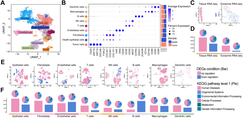
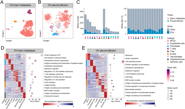
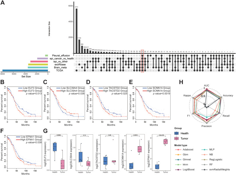
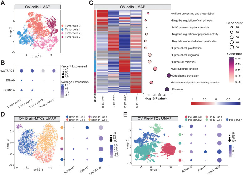
Methods: We integrated single-cell RNA sequencing (scRNA-seq) data from primary tumors and metastatic lesions with bulk tissue transcriptomes and plasma-derived exosomal RNA sequencing (RNA-seq). Differentially expressed genes (DEGs) shared across tumor cells, metastatic subpopulations, and exosomes were identified through intersection analysis. Candidate genes were validated in clinical specimens using qPCR and immunohistochemistry. We then applied ten machine learning algorithm to exosomal transcriptomic data to evaluate diagnostic performance and identify the optimal classifier. Tumor cell differentiation states were evaluated using CytoTRACE, and intercellular communication was analyzed with CellChat.
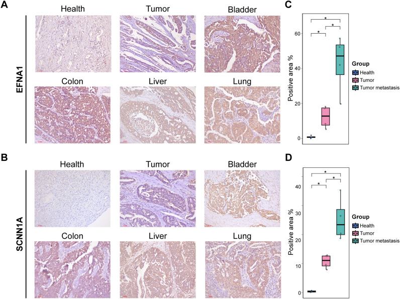
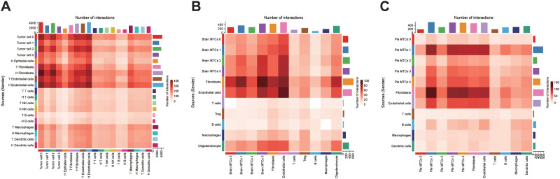
Results: Intersection analysis highlighted 52 overlapping DEGs, of which SCNN1A and EFNA1 emerged as the top prognostic indicators. Both genes were significantly upregulated in tumor tissues, metastatic foci, and plasma exosomes (P < 0.01). The exosome-based Adaboost model had an area under the curve of 0.955 in an independent test cohort. Single-cell subcluster analyses revealed high SCNN1A/EFNA1 expression correlated with stem-like differentiation states and enriched pathways associated with immune evasion and adhesion. CellChat analysis demonstrated that highly differentiated tumor cells extensively engaged with fibroblasts and endothelial cells, implying their role in niche formation.
Conclusions: By coupling single-cell, bulk tissue, and exosomal transcriptomics, we elucidated the key molecular drivers of OV metastasis and established SCNN1A and EFNA1 as promising non-invasive biomarkers. This multi-omics framework provides an effective strategy for early detection and a better understanding of metastatic progression in OV.
Keywords: biomarker; exosome; metastasis; ovarian cancer; single-cell RNA sequencing.
Copyright © 2025 Tang, Pang, Wang, Lin, Chen, Wu and Cui.
Conflict of interest statement
The authors declare that the research was conducted in the absence of any commercial or financial relationships that could be construed as a potential conflict of interest.
Figures
Similar articles
-
Integrated single-cell and transcriptomic analysis of bone marrow-derived metastatic neuroblastoma reveals molecular mechanisms of metabolic reprogramming.Sci Rep. 2025 Aug 5;15(1):28519. doi: 10.1038/s41598-025-13626-8. Sci Rep. 2025. PMID: 40764361 Free PMC article.
-
Identification of blood-derived exosomal tumor RNA signatures as noninvasive diagnostic biomarkers for multi-cancer: a multi-phase, multi-center study.Mol Cancer. 2025 Mar 1;24(1):60. doi: 10.1186/s12943-025-02271-4. Mol Cancer. 2025. PMID: 40025576 Free PMC article.
-
Serum exosomal hsa-let-7f-5p: A potential diagnostic biomarker for metastatic pancreatic cancer detection.World J Gastroenterol. 2025 Jul 14;31(26):109500. doi: 10.3748/wjg.v31.i26.109500. World J Gastroenterol. 2025. PMID: 40678708 Free PMC article.
-
Intraoperative frozen section analysis for the diagnosis of early stage ovarian cancer in suspicious pelvic masses.Cochrane Database Syst Rev. 2016 Mar 1;3(3):CD010360. doi: 10.1002/14651858.CD010360.pub2. Cochrane Database Syst Rev. 2016. PMID: 26930463 Free PMC article.
-
Blood biomarkers for the non-invasive diagnosis of endometriosis.Cochrane Database Syst Rev. 2016 May 1;2016(5):CD012179. doi: 10.1002/14651858.CD012179. Cochrane Database Syst Rev. 2016. PMID: 27132058 Free PMC article.
References
-
- Roett MA, Evans P. Ovarian cancer: an overview. Am Fam Physician. (2009) 80:609–16., PMID: - PubMed
MeSH terms
Substances
LinkOut - more resources
Full Text Sources
Medical
Miscellaneous