

Paracetamol Induces Apoptosis, Reduces Colony Formation, and Increases PTPRO Gene Expression in Human Embryonic Kidney HEK 293 Cells
- PMID: 40787714
- PMCID: PMC12337081
- DOI: 10.1002/jbt.70366
Paracetamol Induces Apoptosis, Reduces Colony Formation, and Increases PTPRO Gene Expression in Human Embryonic Kidney HEK 293 Cells
Abstract
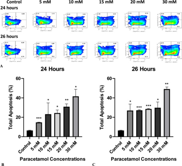
Paracetamol (acetaminophen) is a widely used analgesic and antipyretic drug. Although many studies are available showing that paracetamol causes hepatic damage, few studies exist on renal damage. Protein tyrosine phosphatase receptor type O (PTPRO) protein has tyrosine phosphatase activity and plays a role in the regulation of the renal glomerular pressure/filtration rate relationship in podocyte structure. The PTPRO gene also exhibits characteristics of a candidate tumor suppressor gene. In this study, apoptosis, cell cycle, colony formation, and PTPRO gene expression of the human embryonic kidney (HEK 293) cells were assessed by treating with paracetamol at different concentrations for 24 and 26 h in at least three independent trials. Paracetamol induced dose-dependent apoptosis at all concentrations (5, 10, 15, 20, and 30 mM) for both 24 and 26 h (p < 0.05-p < 0.001). Paracetamol decreased dose-dependent colony-forming potential of cells at 10.000 cells seeding in HEK 293 cells at all concentrations (5, 10, 15, 20, and 30 mM) (p < 0.05-p < 0.001). In HEK 293 cells, paracetamol also increased cell population in the G0-G1 phase at low 5 mM concentration for 26 h (p < 0.05), while reducing cell population in the G2-M phase of the cell cycle at 10 mM and increasing at 30 mM concentration for 24 h (p < 0.05 and p < 0.05, respectively). Paracetamol at high concentration (30 mM) increased PTPRO gene expression in HEK 293 cells for both 24 and 26 h (p < 0.05). In conclusion, our results may indicate that paracetamol may suppress the development of renal cancer by inducing apoptosis and reducing colony formation in a dose-dependent manner, and upregulating PTPRO gene expression in HEK 293 cells at high concentration, in addition to its renal-damaging role. Further research is necessary to determine the potential role or detrimental effects of paracetamol on the development of renal cancer, as the mechanism underlying its effects on renal damage and cancer formation remains unclear.
Keywords: HEK 293 cells; PTPRO; acetaminophen (paracetamol); apoptosis; cell cycle; colony formation; renal cancer.
© 2025 The Author(s). Journal of Biochemical and Molecular Toxicology published by Wiley Periodicals LLC.
Conflict of interest statement
The authors declare no conflicts of interest.
Figures


Similar articles
-
Ibuprofen and/or paracetamol (acetaminophen) for pain relief after surgical removal of lower wisdom teeth.Cochrane Database Syst Rev. 2013 Dec 12;2013(12):CD004624. doi: 10.1002/14651858.CD004624.pub2. Cochrane Database Syst Rev. 2013. PMID: 24338830 Free PMC article.
-
Codeine, alone and with paracetamol (acetaminophen), for cancer pain.Cochrane Database Syst Rev. 2014 Sep 19;2014(9):CD006601. doi: 10.1002/14651858.CD006601.pub4. Cochrane Database Syst Rev. 2014. PMID: 25234029 Free PMC article.
-
Tramadol with or without paracetamol (acetaminophen) for cancer pain.Cochrane Database Syst Rev. 2017 May 16;5(5):CD012508. doi: 10.1002/14651858.CD012508.pub2. Cochrane Database Syst Rev. 2017. PMID: 28510996 Free PMC article.
-
Interventions for paracetamol (acetaminophen) overdose.Cochrane Database Syst Rev. 2018 Feb 23;2(2):CD003328. doi: 10.1002/14651858.CD003328.pub3. Cochrane Database Syst Rev. 2018. PMID: 29473717 Free PMC article.
-
Paracetamol (acetaminophen) or non-steroidal anti-inflammatory drugs, alone or combined, for pain relief in acute otitis media in children.Cochrane Database Syst Rev. 2016 Dec 15;12(12):CD011534. doi: 10.1002/14651858.CD011534.pub2. Cochrane Database Syst Rev. 2016. Update in: Cochrane Database Syst Rev. 2023 Aug 18;8:CD011534. doi: 10.1002/14651858.CD011534.pub3. PMID: 27977844 Free PMC article. Updated.
References
-
- Chidiac A. S., Buckley N. A., Noghrehchi F., and Cairns R., “Paracetamol (Acetaminophen) Overdose and Hepatotoxicity: Mechanism, Treatment, Prevention Measures, and Estimates of Burden of Disease,” Expert Opinion on Drug Metabolism & Toxicology 19, no. 5 (2023): 297–317, 10.1080/17425255.2023.2223959. - DOI - PubMed
-
- Daoud A., Dalhoff K. P., and Petersen T. S., “Paracetamol‐Induced Acute Kidney Injury in the Absence of Acute Liver Injury: A Retrospective Cohort Study,” Toxicology Communications 6, no. 1 (2022): 97–100, 10.1080/24734306.2022.2117941. - DOI
-
- Abdel‐Zaher A. O., Abdel‐Rahman M. M., Hafez M. M., and Omran F. M., “Role of Nitric Oxide and Reduced Glutathione in the Protective Effects of Aminoguanidine, Gadolinium Chloride and Oleanolic Acid Against Acetaminophen‐Induced Hepatic and Renal Damage,” Toxicology 234, no. 1–2 (2007): 124–134, 10.1016/j.tox.2007.02.014. - DOI - PubMed
MeSH terms
Substances
LinkOut - more resources
Full Text Sources
Medical
Miscellaneous
