. 2025 Jan-Dec:34:9636897251362107.
doi: 10.1177/09636897251362107. Epub 2025 Aug 11.

Extracellular matrix protein anosmin-1 regulates Schwann cell-astrocyte interaction for regenerative axon targeting in dorsal root crush injury model

Affiliations

Extracellular matrix protein anosmin-1 regulates Schwann cell-astrocyte interaction for regenerative axon targeting in dorsal root crush injury model

Xichen Xuan et al. Cell Transplant. 2025 Jan-Dec.

Abstract

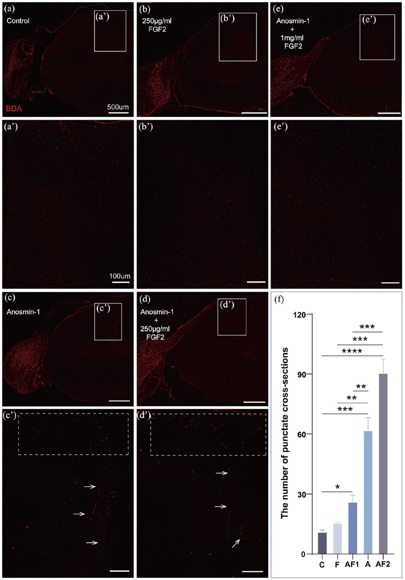
Schwann cell (SC) transplantation is considered as a promising strategy for spinal cord injury. However, SCs show less capability in assisting the regenerative axons to penetrate through astrocyte (AS)-formed scar barrier. Anosmin-1, an extracellular matrix glycosylated adhesion protein expressed in the olfactory bulb, is involved in olfactory ensheathing cells and reborn olfactory nerve axons continually penetrating the glial barrier and targeting the olfactory bulb. In this study, we employ a dorsal root crush injury model treated with anosmin-1. A vertical climbing test was used for behavioral analysis and immunohistochemical study for SC/AS interaction in regenerative axon targeting. Anosmin-1 improved rat forepaw grasping as revealed by forelimb proprioception assessment. After treated with anosmin-1, p75+ immature SCs and P0+ mature SCs mingled well with ASs at the peripheral/central glial interface, reforming the glial barrier from a tight to loose structure. Furthermore, regenerated axons traced by BDA staining revealed proper axonal targeting to the dorsal horn of the spinal cord. These results suggest that anosmin-1 can regulate SC/AS interactions at the peripheral/central boundary site to open the glial barrier for regenerating axons crossing, targeting, and establishing functional neuronal circuits. Anosmin-1 might have a potential application in repair of spinal cord injuries, particularly in combination with SCs for autologous cell transplantation.

Keywords: Schwann cell; anosmin-1; astrocyte; glial boundary; spinal cord injury.


Conflict of interest statement

Declaration of conflicting interestsThe author(s) declared no potential conflicts of interest with respect to the research, authorship, and/or publication of this article.

Figures

Graphical Abstract
Graphical Abstract
Dorsal root crush injury model diagram; rat; pre- and post-injury comparison; axon lesion imaging.
Figure 1.
Dorsal root crush injury model. (a) Experimental design diagram. The dorsal roots of the C6-T1 segments on the left side were individually clamped close to the spinal cord surface, and reagents were injected at the injury site near the spinal cord. For nerve tracing, BDA was injected into the dorsal root ganglion 4 weeks after modeling. (b) Postoperatively, the left forepaw showed a characteristic of fist clenching, which was notably different from the right forepaw (uninjured side). (c) A representative image showing a distinct lesion at the entry zone between the dorsal root ganglion and the spinal cord following the crush injury. 68 kDa-NF staining labeled small-diameter axons. (d, e) High-magnification image of the boxed area in Figure c, the crushed notch is marked with a white arc (horizontal section. Red: 68 kDa-NF; blue: DAPI).
An experiment on human forepaw grasping in a vertical climbing test; success rate and error scores over 42 days post-operation using different treatments.
Figure 2.
Behavioral test—vertical climbing test. (a) Example of normal grasp; (b) error scoring scheme for the left forepaw locating and grasping the grid bars. 0, successful grasp; 1, paw reaches the grid but does not grasp; 2, paw extends to the level of the wrist; 3, to the level of the elbow; 4, to the level of the axilla. Figure b adapted from Minkelyte et al. (c, d) Anosmin-1 improves grasping ability in vertical climbing test. (c) The obvious error rate of forepaw positioning for grasping the bar in the climbing task [time: F(5.521, 209.8) = 167.5, P < 0.0001; group: F(4, 41) = 24.37, P < 0.0001; interaction: F(28, 266) = 5.586, P < 0.0001]. **P < 0.01, ****P < 0.0001, group C versus group AF2; ##P < 0.01, ###P < 0.001, ####P < 0.0001, group C versus group A. (d) Mean error scores for positioning the forepaw to grasp the bar in the climbing task [time: F(3.887, 144.9) = 329.6, P < 0.0001; group: F(4, 40) = 14.49, P < 0.0001; interaction: F(28, 261) = 4.141, P < 0.0001]. ***P < 0.001, ****P < 0.0001, group C versus group AF2; #P < 0.05, ##P < 0.01, group C versus group A. Groups C, A, and AF2 (n = 10); groups F and AF1 (n = 7). Scores during the 6-week test period. Mean ± standard error. Two-way ANOVA.
This image shows the effects of various dosages of FGF2 on the migration and interaction of Anosmin-1 with mature satellite cells (SCs) and adipose stem cells (ASs) in a spinal cord environment, with high-magnification images of cell interactions and a bar graph comparing the farthest migration distances of P0+ SCs across different groups.
Figure 3.
Anosmin-1 modulates mature SCs and ASs interaction in the peripheral/central boundary site. Tight boundary formation between mature SCs and ASs at the peripheral and central interface in group C (a, a’) and group F (b, b’); (c, c’) group A. Mature SCs were seen in two consecutive segments near the central border, with striated AS in the peripheral side; (d, d’) group AF2. Several clusters of mature SCs were observed at the peripheral-central junction, and the AS in the PNS migrated farther than in group A; (e, e’) group AF1. Scattered mature SCs were present in the spinal cord, while ASs migration to the peripheral side was observed. (a’, b’, c’, d’, e’) High-magnification images within the white boxes in the corresponding figures. (f) Quantitative analysis showed that the farthest migration distance of P0+ SCs inside the spinal cord was statistically much further in groups A and AF2 as compared with group C and there was no significant difference between groups AF1 and AF2. *P < 0.05, **P < 0.01, ***P < 0.001, ****P < 0.0001. Groups C, A, and AF2 (n = 6); groups F and AF1 (n = 4). Mean ± standard error. One-way ANOVA (coronal section. “*” indicates the side of the spinal cord. Red: P0; Green: GFAP; Blue: DAPI).
The chart displays the results of a study on the interaction between peripheral and central glial cells, using Anosmin-1 and other factors.
Figure 4.
Anosmin-1 modulates immature SCs and ASs interaction in the peripheral/central boundary site. (a, a’) Group C. No interaction was observed between peripheral and central glial cells; (b, b’) group F. No interaction was observed between peripheral and central glial cells; (c, c’) group A. A large number of immature SCs were present centrally near the spinal cord, together with a sparse AS border; (d, d’) group AF2. Rounded and spindle-shaped immature SCs were observed inside the spinal cord, with scattered AS in the peripheral side; (e, e’) group AF1. Scattered immature SCs were seen in the spinal cord, with some ASs in the peripheral side; (a’, b’, c’, d’, e’) High-magnification images within the white boxes in the corresponding figures. (f) Quantitative analysis showed that the farthest migration distance of p75+ SCs inside the spinal cord was statistically much further in groups A and AF2 as compared with group C and there was a significant difference between groups AF1 and AF2. *P < 0.05, **P < 0.01. Groups C, A, and AF2 (n = 6); groups F and AF1 (n = 4). Mean ± standard error. One-way ANOVA (coronal section. “*” indicates the side of the spinal cord. Red: p75; Green: GFAP; Blue: DAPI).
Create a PDF of this image
Figure 5.
Anosmin-1 promotes regenerative axon targeting to the dorsal horn of the spinal cord. In the control group (a) and FGF2 treatment group (b), few of BDA-positive staining was observed in the spinal cord; (c) and (c’) After treated with anosmin-1, the filamentous regenerating axons of varying lengths were observed within the dorsal horn (arrows), and multiple punctates representing cross-sectioned regenerative axon bundles were seen within the dorsal column (dashed box); (d) and (d’) After treated with anosmin-1+250 µg/ml FGF2, more punctates of regenerating axon bundles by cross-section were observed in the dorsal column (dashed box), along with thread-like regenerating axons of varying lengths in the dorsal horn (arrows); (e) After treated with anosmin-1+1 mg/ml FGF2, few of BDA-positive staining was observed in the spinal cord; (f) Quantitative analysis showed that the number of punctates in the dorsal column in groups A, AF1 and AF2 were higher than that in group C and much more punctates were present in groups A, AF2 as compared with AF1. *P < 0.05, **P < 0.01, ***P < 0.001, ****P < 0.0001. Groups C, A, and AF2 (n = 4); groups F and AF1 (n = 3). Mean ± standard error. One-way ANOVA. (a’, b’, c’, d’, e’) High-magnification images of the regions within the white boxes in the corresponding figures (horizontal section; red: BDA).
Diffused distribution of centrally located SCs without wrapping regenerating axons. (a) Horizontal section of the spinal cord and connected dorsal root nerves, showing the diffused distribution of central p75+ cells on the left side as compared with the right side; (A1) High-magnification image of the boxed area in Fig. 6a, showing the irregularly scattered distribution of SCs; (A2) High-magnification image of the boxed area in Fig. 6A1. SCs (arrows) did not wrap regenerating axons (horizontal section; red: BDA; green: p75; blue: DAPI).
Figure 6.
Diffused distribution of centrally located SCs without wrapping regenerating axons. (a) Horizontal section of the spinal cord and connected dorsal root nerves, showing the diffused distribution of central p75+ cells on the left side as compared with the right side; (a1) High-magnification image of the boxed area in Fig. 6a, showing the irregularly scattered distribution of SCs; (a2) High-magnification image of the boxed area in Fig. 6a1. SCs (arrows) did not wrap regenerating axons (horizontal section; red: BDA; green: p75; blue: DAPI).

Similar articles

References

    1. Zhou H, Lou Y, Chen L, Kang Y, Liu L, Cai Z, Anderson DB, Wang W, Zhang C, Wang J, Ning G, et al. Epidemiological and clinical features, treatment status, and economic burden of traumatic spinal cord injury in China: a hospital-based retrospective study. Neural Regen Res. 2024;19(5):1126–33. doi: 10.4103/1673-5374.382257. - DOI - PMC - PubMed
    1. Clifford T, Finkel Z, Rodriguez B, Joseph A, Cai L. Current advancements in spinal cord injury research-glial scar formation and neural regeneration. Cells. 2023;12(6):853. doi: 10.3390/cells12060853. - DOI - PMC - PubMed
    1. Tran AP, Warren PM, Silver J. The biology of regeneration failure and success after spinal cord injury. Physiological Reviews. 2018;98(2):881–917. doi: 10.1152/physrev.00017.2017. - DOI - PMC - PubMed
    1. Gómez RM, Sánchez MY, Portela-Lomba M, Ghotme K, Barreto GE, Sierra J, Moreno-Flores MT. Cell therapy for spinal cord injury with olfactory ensheathing glia cells (OECs). Glia. 2018;66(7):1267–301. doi: 10.1002/glia.23282. - DOI - PubMed
    1. Li BC, Xu C, Zhang JY, Li Y, Duan ZX. Differing Schwann cells and olfactory ensheathing cells behaviors, from interacting with astrocyte, produce similar improvements in contused rat spinal cord’s motor function. J Mol Neurosci. 2012;48(1):35–44. doi: 10.1007/s12031-012-9740-6. - DOI - PubMed

MeSH terms

Substances