. 2025 Aug 27;13(34):10621-10634.
doi: 10.1039/d5tb00816f.

End group chemistry modulates physical properties and biomolecule release from biodegradable polyesters

Affiliations

End group chemistry modulates physical properties and biomolecule release from biodegradable polyesters

Matthew A Borrelli et al. J Mater Chem B. .

Abstract

Long-acting injectable protein therapeutics are a rapidly advancing arm of pharmaceuticals. A promising and versatile class of such formulations involves encapsulation of therapeutic protein within poly(lactic-co-glycolic acid) (PLGA) degradable microparticles (MP) to shield the protein from enzymatic degradation and control the release rate. However, models based on degradation and erosion of PLGA polymer matrices do not always fully capture release behavior, due in part to electrostatic interactions between the polymer terminal group and encapsulated compound. The repertoire of functionalized PLGA polymers commercially available has now expanded to include terminal group chemistries that may significantly alter polymer characteristics including charge, hydrophobicity, and erosion. This work aims to explore how PLGA terminal group chemistry affects polymer physical properties and charged biomolecule release kinetics. PLGA with hydroxyl (PLGA-OH), amine (PLGA-NH2), or carboxylic acid (PLGA-COOH) terminal groups that have neutral, positive, or negative charge, respectively, were evaluated. Experiments assessing the physical properties of the polymers indicate PLGA-NH2 has reduced hydrophobicity, degrades faster, exhibits emulsion stabilizing behavior, and has reduced phagocytic clearance by bone marrow derived macrophages. Charged biomolecule release rates are increased from PLGA-NH2 MPs and slightly accelerated from PLGA-OH MPs, compared to PLGA-COOH MPs. These studies provide further insight into the interactions between charged biomolecules and the encapsulating polymer and could provide additional tools to tune release for various protein therapeutics that experience such interactions.


Conflict of interest statement

There are no conflicts to declare.

Figures

Fig. 1
Fig. 1. Amine functionalized PLGA displays altered physical properties. PLGA polymers terminated with carboxylic acid (–COOH), amine (–NH2), and hydroxyl (–OH) groups were characterized. (A) Representative images and (B) quantitative analysis of water droplet contact angle on polymer films shows PLGA–NH2 reduces the surface tension (contact angle) of water (N = 30). (C) Representative DSC analysis for −20 to 150 °C at 10 °C min−1 ramp for each of the PLGA polymers is shown, in which the circle denotes the glass transition temperature (Tg). (D) The measured Tg from N = 3 independent experiments shows reduced Tg for PLGA–NH2. Statistical comparisons were made using one-way ANOVA followed by Šídák's multiple comparisons test ***p ≤ 0.001, ****p ≤ 0.0001.
Fig. 2
Fig. 2. Amine-terminated PLGA exhibits emulsion stabilization and osmotic behavior during microparticle formulation. A series of experiments were conducted to determine how PLGA end-caps impact MP formation during double emulsion formulation procedures. (A) Initial evaluation of microparticles (MP) formulated with deionized water for the inner aqueous phase (non-porousMP) shows increased pore formation with PLGA–NH2 microparticles. Scale bar is 10 μm, 1000× magnification. (B) PLGA was incubated in deionized water for 8 h to allow any low Mw polymer fragments to dissolve. Measured osmolarity of the supernatant shows PLGA–NH2 has greater osmolarity than then other PLGA polymers. (C) Representative cross sections of microparticles formulated with an inner aqueous phase osmolarity of 15 mOsm (porousMP). Scale bar is 5 μm, 2000× magnification. (D) Inner occlusion diameter and (E) occlusion density was determined by analyzing the SEM images (C) in QuPath. Inner occlusion diameter is reduced for PLGA–NH2 MPs and increased for PLGA–OH MPs, relative to PLGA–COOH MPs, while the density of occlusions within PLGA–NH2 microparticles is increased. Results represent the average distribution of inner occlusions for N = 4 to 6 microparticle cross sections. Statistical comparisons were made using one-way ANOVA followed by Šídák's multiple comparisons test *p ≤ 0.05, **p ≤ 0.01, ****p ≤ 0.0001.
Fig. 3
Fig. 3. The rate of degradation and evolution of intraparticle pH is accelerated in amine functionalized PLGA. Porous microparticles formulated with end-capped PLGA were incubated in HEPES buffer to undergo degradation by hydrolysis. (A) Measurement of supernatant and intraparticle pH shows differential rates of acidification among the polymers. Notably, intraparticle pH rapidly reduces in PLGA–NH2, but the supernatant lags unlike the other end-capped PLGA. (B) Size exclusion chromatography data shows number averaged molecular weight reduces faster for PLGA–NH2. (C) log transformed SEC data was fitted to a linear degradation model (log10(%Deg.) = −k*t) for (1) non-catalytic erosion and (2) acid-catalyzed erosion phases of degradation. Degradation constants are in Table 1. Data represent mean ± sd for N = 3 samples. Statistical comparisons were made using two-way ANOVA followed by Tukey's multiple comparisons test. Comparisons shown in (A) are for PLGA–NH2 relative to PLGA–COOH. **p ≤ 0.01, ****p ≤ 0.0001.
Fig. 4
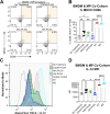
Fig. 4. End-cap PLGA groups augment inflammatory macrophage differentiation and hydroxyl terminal groups are less immunogenic. Murine BMDMΦs and PLGA MPs were co-cultured for 3 days and analyzed by flow cytometry. Size distributions of MPs used for these assays are reported in Fig. S2B. (A) Flow plots of live CD11b+ F4/80+ BMDMΦs expressing MHCII and CD86. (B) Graph depicts frequency of double positive populations. (C) Histograms of IL-12 fluorescence and (D) calculated median fluorescence intensity (MFI). Data represents mean ± sd for N = 9 samples (n = 3 wells per mouse). Statistical comparisons were made using one-way ANOVA followed by Tukey's multiple comparisons test. *p ≤ 0.05, **p ≤ 0.01, ***p ≤ 0.001, ****p ≤ 0.0001.
Fig. 5
Fig. 5. PLGA–NH2 microparticles exhibit reduced internalization by macrophages. Murine BMDMΦs and PLGA MPs were co-cultured for 24 hours and analyzed by ImageStream or spectral cytometry. (A) Microparticle size distribution and (B) zeta potential for microparticles formulated to contain Alexa fluor 680 labeled dextran. (C) ImageStream calculated internalization score, which is based on a ratio of the AF680 probe (MP) to cellular stain (CD11b) intensity. Flow cytometric quantification of (D) M1 phenotypic marker iNOS and (E) M2 phenotypic marker CD206 for BMDMΦs with internalized microparticles (+) as compared to BMDMΦs without internalized microparticles (−). Data in (A) represents the size distribution of 10 000 events for each formulation. Data in (B–E) represents the mean ± SD for N = 3 individual experiments. Data in (C) represent pooled data from N = 3 individual experiments. Statistical comparisons were made using one-way ANOVA followed by Tukey's multiple comparisons test. **p ≤ 0.01, ****p ≤ 0.0001.
Fig. 6
Fig. 6. Five key amino acids are responsible for biomolecule net charge. Biomolecules encapsulated in PLGA MPs have potential to exhibit charge interactions with the polymer terminal groups. (A) Calculations of amino acid (AA) residue charge across typical supernatant and intraparticle pH ranges show negatively charged residues become uncharged in acidic conditions. Three biomolecules were selected for encapsulation: 2 peptides with positive and neutral charge and Mw ∼ 2.5 kDa and a more complex protein, CCL22, Mw ∼ 10 kDa. The sequences for these biomolecules are detailed in (B), and residues with potential for positive (blue) or negative (purple) charge are highlighted for each. (C) Net charge calculations were performed for these biomolecules from pH 0 to 7.
Fig. 7
Fig. 7. Biomolecule release & encapsulation with end-cap PLGA shows charge-dependent behavior. Cumulative release of positively and neutrally charged peptide from porous MPs with altered PLGA end caps was quantified. (A) Size distributions of microparticles loaded with the neutral peptide, positive peptide, and CCL22 protein. Table S2 details the mean and standard deviation of the size distribution for each formulation. (B) Neutral peptide release kinetics demonstrate biomolecule release in the absence of charge interactions. (C) Positively charged peptide release is also influenced by peptide–polymer electrostatic interactions. (D) For the larger and more complex protein, CCL22, the order of release is consistent with that for the positive peptide, but release from PLGA–OH and PLGA–COOH MPs is significantly depressed compared to PLGA–NH2 MPs. Data in A represent volume impedance measurement of particle size for N = 10 000 events. Data in (B)–(D) represent mean + sd for N = 3 independent release experiments.

Similar articles

References

    1. Global Markets and Manufacturing Technologies for Protein Drugs, BCC Research, 2021
    1. Werle M. Bernkop-Schnurch A. Amino Acids. 2006;30:351–367. doi: 10.1007/s00726-005-0289-3. - DOI - PubMed
    1. Gandell D. L. Bienen E. J. Gudeman J. Patient Prefer. Adherence. 2019;13:351–361. doi: 10.2147/PPA.S187120. - DOI - PMC - PubMed
    1. Gonella A. Grizot S. Liu F. Lopez Noriega A. Richard J. Expert Opin. Drug Delivery. 2022;19:927–944. doi: 10.1080/17425247.2022.2105318. - DOI - PubMed
    1. Makadia H. K. Siegel S. J. Polymers. 2011;3:1377–1397. doi: 10.3390/polym3031377. - DOI - PMC - PubMed

Substances

LinkOut - more resources