. 2025 Aug 11;18(1):75.
doi: 10.1186/s12284-025-00830-3.

Salinity Stress Induces Phase Separation of Plant BARENTSZ to Form Condensates

Affiliations

Salinity Stress Induces Phase Separation of Plant BARENTSZ to Form Condensates

Peigang Li et al. Rice (N Y). .

Abstract

Phase separation (PS) of BARENTSZ (BTZ), a core member of the exon-junction complex (EJC), is involved in various physiological and developmental processes in animals. However, less is known about plant equivalents. Here, we demonstrated that the loss of function of Oryza sativa BTZ genes (OsBTZs) reduced rice tolerance to salinity stress. Moreover, OsBTZ proteins underwent PS independent of other core members of EJC, forming condensates under salt stress. OsBTZs may recruit proteins that play roles in the salt tolerance response to form cytoplasmic condensates, which act as stress granules. The predicted prion-like domain (PrLD), that originated ancestrally and is functionally conserved, was demonstrated to be key to the PS of OsBTZs upon NaCl treatment. This work revealed a new role for plant BTZs through an evolutionarily conserved mechanism-PS-in the formation of condensates in response to salinity stress, thus providing new insights into the adaptive evolution of plant BTZs under abiotic stress.

Keywords: BARENTSZ (BTZ); Evolution; Phase separation; Rice; Salt stress; Stress granules.


Conflict of interest statement

Declarations. Ethics Approval and Consent to Participate: Not applicable. Consent for Publication: Not applicable. Competing Interests: The authors declare no competing interests.

Figures
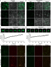
Fig. 1
Fig. 1
Condensations of OsBTZs after NaCl treatment. a, b Subcellular localization of OsBTZ1-GFP and OsBTZ3-GFP. Transient expression was performed in the leaf cells of Nicotiana benthamiana under untreated and 200 mM NaCl conditions. GFP, green fluorescent protein; Bright, bright field; Merged, merged image of GFP and Bright. Bars = 100 μm. c, d Images and quantification of fluorescence recovery after photobleaching (FRAP) of OsBTZ1- (c) and OsBTZ3-containing (d) granules, respectively. The images shown are representative of independent observations of OsBTZ1-GFP and OsBTZ3-GFP. Plots on the bottom indicate quantified fluorescent signals over the time course after photobleaching. e Colocalization of OsBTZ1-GFP and OsBTZ3-GFP with ANGUSTIFOLIA (AN) in untreated and 200 mM NaCl-treated samples. These fusion proteins were transiently expressed in the leaf cells of N. benthamiana. Merged, merged image of GFP and mCherry. Bars = 200 μm
Fig. 2
Fig. 2
Condensation by OsBTZs is dependent on the presence of prion-like domain (PrLD). a, b Protein domain prediction of OsBTZ1 (a) and OsBTZ3 (b) via PLAAC (http://plaac.wi.mit.edu). PrLD, prion-like domain; NLS, nuclear localization sequence; SELOR, speckle localizer and RNA binding domain. c, d Protein domain structure and various truncated versions of OsBTZ1 (c) and OsBTZ3 (d) as indicated. △PrLD, removing PrLD; △SELOR, removing SELOR; △SELOR/PrLD, removing both SELOR and PrLD. e, Expression and subcellular localization of green fluorescent protein (GFP)-tagged truncated OsBTZs, as shown in (c, d). Condensate formation assays were performed in the leaf cells of N. benthamiana under NaCl treatments. Bars = 100 μm
Fig. 3
Fig. 3
Protein components of OsBTZ-associated condensates. a Venn diagram showing the potential proteins that interact with OsBTZ1 and OsBTZ3 under normal and 200 mM NaCl conditions. The total number of candidate interacting proteins obtained via library screening via yeast two-hybrid analysis is summarized below. b Gene Ontology (GO) analysis of proteins that interact with OsBTZ1 and OsBTZ3. c Bimolecular florescence complementation (BiFC) analysis of the protein‒protein interactions between OsBTZs and two potential candidate proteins in the leaf cells of Nicotiana benthamiana. The alteration of the subcellular localization of the interacting signal under 200 mM NaCl conditions relative to that under normal (untreated) conditions indicates the formation of condensates. YFP, yellow fluorescent protein; Bright, bright field; Merged, merged YFP and Bright. Bars = 100 μm
Fig. 4
Fig. 4
OsBTZs confer tolerance to salt stress in rice. a Seed germination of the osbtz1 mutants (L1 and L3), osbtz3 mutants (L1 and L2), and osbtz1/3 mutant compared with their corresponding wild-type (WT) ‘ZH10’ under 200 mM NaCl stress for 6 days. Bars = 3.6 cm in the upper part, and bars = 1 cm in the lower part. b Seed germination rates recorded from day 1 to day 6 under both normal conditions and 200 mM NaCl treatments. Each germination experiment was conducted with three biological replicates (50 seeds per replicate), and the means are presented. P values were estimated on day 6 for all genotypes relative to the WT (P1) and single osbtz mutants relative to the double osbtz1/3 mutant (P2) via a two-tailed Student’s t test. c Seed setting rates of WT, osbtz1-L1, osbtz1-L3, osbtz3-L1, osbtz3-L2, and osbtz1/3 under both normal conditions and 200 mM NaCl treatments. The means and standard deviations are presented (n = 10). The lowercase letters above columns indicate significant differences (P < 0.05) in one-way analysis of variance determined by SPSS software 24.0 (IBM Corp, NY, USA)
Fig. 5
Fig. 5
Evolutionary analysis of PrLD and BTZ phase separation. a Maximum likelihood (ML) tree of the BTZs in selected representative species from plants. The predicted prion-like domain (PrLD) of these BTZ proteins are listed on the right. A total of 19 BTZ genes from 11 plant species are displayed, including MpBTZ (Marchantia polymorpha), CpBTZ (Ceratodon purpureus), PpBTZ (Physcomitrella patens), NcBTZ (Nymphaea colorata), OsBTZ (Oryza sativa Japonica), SvBTZ (Setaria viridis), CarBTZ (Capsella rubella), AtBTZ (Arabidopsis thaliana), SlBTZ (Solanum lycopersicum), NaBTZ (Nicotiana attenuata), and PfBTZ (Physalis floridana). b ML tree of the BTZs from selected representative animal species. The stick figure of each chosen animal and the predicted PrLDs are listed. BTZ genes from 22 animal species of 12 families are displayed, including HcoBTZ (Hemonchus contortus), SmaBTZ (Schistosoma mansoni), AruBTZ (Asterias rubens), PmaBTZ (Pecten maximus), DmeBTZ (Drosophila melanogaster), AmeBTZ (Apis mellifera), CseBTZ (Cryptotermes secundus), EsiBTZ (Eriocheir sinensis), HmaBTZ (Homarus americanus), StrBTZ (Silurana tropicalis), DreBTZ (Danio rerio), TniBTZ (Tetraodon nigroviridis), TruBTZ (Takifugu rubripes), CmiBTZ (Callorhinchus milii), CtoBTZ (Chauna torquata), EgaBTZ (Egretta garzetta), NnaBTZ (Naja naja), MreBTZ (Mauremys reevesii), MmuBTZ (Mus musculus), LafBTZ (Loxodonta africana), CfaBTZ (Canis lupus familiaris), and HsaBTZ (Homo sapiens). In a and b, the MpBTZ from Marchantia polymorpha was used as an outgroup. Bootstrap values ≥ 50 are presented on the corresponding node of the phylogenetic tree, as are the ancestral state of the PrLD was reconstructed and highlighted in pies, and blue and red indicate the presence and absence of PrLD, respectively. SELOR, speckle localizer, and RNA binding domain. c Subcellular localization of nine BTZs from six plant species under untreated (normal) and 200 mM NaCl conditions. The fusion protein BTZ–GFP, as indicated, was transiently expressed in the leaf cells of Nicotiana benthamiana. GFP, green fluorescent protein. Bars = 150 μm

Similar articles

References

    1. Ali A, Petrov V, Yun DJ, Gechev T (2023) Revisiting plant salt tolerance: novel components of the SOS pathway. Trends Plant Sci 28:1060–1069 - PubMed
    1. Asthana S, Martin H, Rupkey J, Patel S, Yoon J, Keegan A, Mao Y (2022) The physiological roles of the exon junction complex in development and diseases. Cells 11:1192 - PMC - PubMed
    1. Baguet A, Degot S, Cougot N, Bertrand E, Chenard MP, Wendling C, Kessler P, Le Hir H, Rio MC, Tomasetto C (2007) The exon-junction-complex-component metastatic lymph node 51 functions in stress-granule assembly. J Cell Sci 120:2774–2784 - PubMed
    1. Ballut L, Marchadier B, Baguet A, Tomasetto C, Séraphin B, Le Hir H (2005) The exon junction core complex is locked onto RNA by inhibition of eIF4AIII ATPase activity. Nat Struct Mol Biol 12:861–869 - PubMed
    1. Bartkowska K, Tepper B, Turlejski K, Djavadian RL (2018) Roles of the exon junction complex components in the central nervous system: a mini review. Rev Neurosci 29:817–824 - PubMed

LinkOut - more resources