

Sloping retina: a novel feature associated with primary open-angle glaucoma in an African ancestry cohort
- PMID: 40789618
- PMCID: PMC12336614
- DOI: 10.1136/bmjophth-2025-002224
Sloping retina: a novel feature associated with primary open-angle glaucoma in an African ancestry cohort
Abstract
Objective: To define sloping of the retina, a novel stereoscopic feature in primary open-angle glaucoma (POAG), and to evaluate its prevalence and associated risk factors in an African ancestry population.
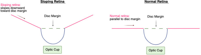
Methods and analysis: Digital stereo disc images were graded for sloping by trained non-physician graders. We defined a sloping retina as one that slanted downward towards the disc margin instead of existing on the same plane as the disc margin. A 'sloping retina' approached the disc margin at an angle along at least one-third of the disc's circumference. The ocular and demographic risk factors of sloping were evaluated by univariable and multivariable logistic regression models.
Results: The prevalence of sloping in eyes with POAG was 22.0% (95% CI 20.6% to 23.4%). In a multivariable analysis, compared with eyes without sloping, eyes with sloping were less likely to have disc haemorrhages (p=0.03) and more likely to have a tilted disc (p<0.001), larger cup-to-disc ratio ((defined as 0.7-1), p=0.002), grey crescent (p=0.02), nasalisation of the vessels (p=0.01), moderate or deep cup depth (p<0.001) and conical cup shape (p<0.001). Sloping was not associated with any demographic characteristics in the multivariable analysis.
Conclusion: Associated with risk factors of advanced POAG, sloping presents as a novel feature that warrants further study to determine its mechanisms of development and prevalence in other study populations. Study limitations include: large difference in the number of eyes with and without sloping, potential morphological expressions of other phenotypes posing as sloping, impact of anatomical variability on grading, inherent biases when grading stereoscopic images and absence of a control or glaucoma suspect group. Future research into this phenotype in POAG patients might determine whether sloping retina is the result of or a precursor to glaucomatous damage, leading to a better understanding of POAG.
Keywords: Glaucoma; Imaging; Retina.
© Author(s) (or their employer(s)) 2025. Re-use permitted under CC BY-NC. No commercial re-use. See rights and permissions. Published by BMJ Group.
Conflict of interest statement
Competing interests: None declared.
Figures
Similar articles
-
Optic nerve head and fibre layer imaging for diagnosing glaucoma.Cochrane Database Syst Rev. 2015 Nov 30;2015(11):CD008803. doi: 10.1002/14651858.CD008803.pub2. Cochrane Database Syst Rev. 2015. PMID: 26618332 Free PMC article.
-
A comparison of optic disc size and retinal thickness between a glaucomatous and nonglaucomatous African population.Optom Vis Sci. 2025 Jul 1;102(7):436-451. doi: 10.1097/OPX.0000000000002268. Epub 2025 Jun 5. Optom Vis Sci. 2025. PMID: 40478745
-
Comprehensive assessment of glaucoma in patients with high myopia: a systematic review and meta-analysis with a discussion of structural and functional imaging modalities.Int Ophthalmol. 2024 Oct 11;44(1):405. doi: 10.1007/s10792-024-03321-4. Int Ophthalmol. 2024. PMID: 39392516 Free PMC article.
-
Comparison of Peripapillary Choroidal Thickness Between Primary Open-angle Glaucoma, Normal Tension Glaucoma, and Normal Eyes: A Systematic Review and Meta-analysis.Ophthalmol Glaucoma. 2024 Jul-Aug;7(4):359-371. doi: 10.1016/j.ogla.2024.02.008. Epub 2024 Feb 23. Ophthalmol Glaucoma. 2024. PMID: 38403265
-
Peripheral iridotomy for pigmentary glaucoma.Cochrane Database Syst Rev. 2016 Feb 12;2(2):CD005655. doi: 10.1002/14651858.CD005655.pub2. Cochrane Database Syst Rev. 2016. PMID: 26871761 Free PMC article.
References
MeSH terms
LinkOut - more resources
Full Text Sources