. 2025 Aug 11:a041688.
doi: 10.1101/cshperspect.a041688. Online ahead of print.

Telomere Crisis Shapes Cancer Evolution

Affiliations

Telomere Crisis Shapes Cancer Evolution

Joe Nassour et al. Cold Spring Harb Perspect Biol. .

Abstract

Somatic mutations arise in normal tissues and precursor lesions, often targeting cancer-driver genes involved in cell cycle regulation. Most checkpoint-mutant clones, however, remain dormant throughout an individual's lifetime and seldom progress to malignancy, implying the presence of protective mechanisms that limit their expansion and malignant transformation. One such safeguard is telomere crisis-a potent tumor-suppressive barrier that eliminates cells lacking functional checkpoints and evading p53- and pRb-mediated surveillance. While the genomic instability unleashed during telomere crisis can drive clonal evolution, cell death is typically the dominant outcome, with only a rare subset of cells escaping elimination to initiate malignancy. Recognizing the dual role of telomere crisis-suppressing tumor initiation while enabling clonal evolution-is essential for understanding early cancer development and designing strategies to eliminate tumor-initiating cells.


Figures

Figure 1.
Figure 1.
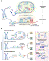
Telomere shortening imposes proliferative barriers and drives clonal evolution. Cancer arises through clonal evolution, with genetic variation providing the substrate for selection. Analyses of intratumoral heterogeneity (ITH) have yielded insights into how evolutionary forces govern tumor growth and progression. Central to this framework is cellular fitness—the ability of a tumor cell to survive, proliferate, and transmit its genotype. Increases in fitness promote clonal expansion and can lead to selective sweeps that favor dominant clones. In the early stages of tumorigenesis, telomere shortening imposes two distinct proliferative barriers: replicative senescence (M1) and crisis (M2). Senescence occurs when critically short telomeres trigger a DNA damage response (DDR), arresting cell division. Inactivation of p53 and retinoblastoma protein ( pRb) allows cells to bypass this checkpoint, enabling continued proliferation and progressive telomere dysfunction. However, these checkpoint-deficient cells eventually reach crisis—a state marked by severe chromosomal instability (CIN) and widespread cell death. Although telomere crisis serves as a powerful tumor-suppressive barrier, the genomic instability it induces also accelerates clonal evolution by generating a broad spectrum of genetic alterations. Within this selective landscape, rare clones that resist cell death, suppress DDR signaling, or restore telomere maintenance—via telomerase reactivation or the alternative lengthening of telomeres (ALT) pathway—can escape crisis and drive malignant progression. Telomere crisis functions as an evolutionary bottleneck, eliminating most precancerous clones while selecting for resilient subpopulations that fuel tumor evolution. (M1) Mortality stage 1, (M2) mortality stage 2.
Figure 2.
Figure 2.
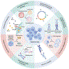
A three-state model of chromosome-end protection. (A) Graphical representation of human telomeres in linear and T-loop configurations. Telomeres distinguish naturally occurring chromosome ends from DNA double-strand breaks, preventing inappropriate repair events and the formation of dicentric chromosomes. They typically comprise several kilobases of repeated 5′-TTAGGG-3′ sequences, ending in a single-stranded G-rich 3′ overhang of 50–300 nt. These repeats are bound by the six-subunit shelterin complex (telomeric repeat-binding factor 1 [TRF1], TRF2, RAP1, TRF1-interacting nuclear factor 2 [TIN2], TPP1, and protection of telomeres 1 [POT1]). Telomeres can form a protective T-loop through invasion of the 3′ overhang into a double-stranded region of telomeric DNA, thereby avoiding recognition by DNA damage response (DDR) machinery. Shelterin represses key DDR enzymes (ataxia-telangiectasia mutated [ATM], ataxia telangiectasia and Rad3-related [ATR], and poly-ADP-ribose polymerase 1 [PARP1]) and blocks the double-strand break (DSB) repair pathways (classical nonhomologous end joining [c-NHEJ], alternative nonhomologous end joining [alt-NHEJ]/micro-homology-mediated end joining [MMEJ], and homologous recombination [HR]). It also prevents excessive DNA end resection at telomeres, preserving genomic stability. (B) Graphical representation of human telomeres in closed-state, intermediate-state, and uncapped-state conformations, as suggested to occur during replicative aging or upon experimental disruption of telomeric repeat-binding factor 2 (TRF2). The intermediate state retains partial shelterin binding, limiting end joining despite DDR signaling; the uncapped state is DDR-positive and highly fusogenic due to insufficient shelterin coverage. Short, dysfunctional telomeres can fuse either with other short telomeres or with nontelomeric genomic loci, yielding dicentric chromosomes. Subsequent breakage of these dicentric chromosomes during cell division initiates breakage–fusion–bridge (BFB) cycles, leading to widespread genomic rearrangements. (RAP1) Repressor activator protein 1, (T-loop) telomere-loop.
Figure 3.
Figure 3.
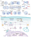
Hallmarks of telomere crisis. In the absence of functional cell cycle checkpoints, continued proliferation beyond the senescence barrier leads to telomere crisis, marked by the loss of end-capping function. Telomeres erode to the point where they are nearly devoid of repeats and can no longer be protected by the shelterin complex. These uncapped chromosome ends are recognized as DNA breaks and processed primarily through microhomology-mediated end joining (MMEJ), producing dicentric chromosomes and initiating breakage–fusion–bridge (BFB) cycles. This process drives widespread genomic instability, including amplifications, loss of heterozygosity, and translocations. The spectrum of telomere-induced genome alterations has recently expanded to include chromothripsis—a catastrophic form of chromosomal rearrangement observed in cancers and some congenital disorders. During crisis, cell death progressively increases until it surpasses proliferation, leading to culture collapse. This death response is triggered by the cytoplasmic accumulation of self-derived nucleic acids, which are sensed as damage-associated molecular patterns by two innate immune pathways. The cyclic GMP–AMP synthase (cGAS)– stimulator of interferon (IFN) genes (STING) axis is activated by cytoplasmic DNA, whereas the Z-DNA-binding protein 1 (ZBP1)–mitochondrial antiviral-signaling protein (MAVS) pathway responds to cytoplasmic telomeric repeat–containing RNA (TERRA) transcripts derived from damaged telomeres. These pathways converge to induce type I IFN signaling and sustained inflammation, culminating in a specialized form of autophagy that eliminates cells in crisis. (M2) Mortality stage 2, (DSB) double-strand break.
Figure 4.
Figure 4.
Telomere-associated molecular patterns and innate immunity. (A) Interchromosomal fusions typically produce dicentric chromosomes carrying two centromeres, whereas intrachromosomal fusions generate pseudo-dicentric chromosomes joined by fused ends. During anaphase, opposing spindle forces pull dicentric chromosomes apart, creating DNA bridges that can be resolved through multiple mechanisms. In one model, actomyosin-driven mechanical force alone can break these bridges, causing simple or extensive chromosome fragmentation. An alternative model posits that the 3′-to-5′ exonuclease three-prime repair exonuclease 1 (TREX1) mediates bridge resolution, requiring transient nuclear envelope breakdown. These processes often yield acentric DNA fragments, which either localize to micronuclei (MN) or are released into the cytosol. In some instances, dicentric chromosomes detach from spindle poles instead of breaking, resulting in lagging chromosomes that can be sequestered into MN. (B) TERRA is a long noncoding RNA transcribed by RNA polymerase II from subtelomeric regions on the C-rich telomeric strand. Polyadenylated telomeric repeat–containing RNA (TERRA) accumulates in the nucleoplasm, whereas nonpolyadenylated TERRA remains associated with telomeric chromatin. The 3′ terminus of TERRA facilitates interactions with telomere-bound proteins, driving the formation of R-loops. TERRA transcripts are also detected in the cytoplasm, where they may be packaged into exosomal vesicles. In the cytoplasm, TERRA can exist as single-stranded RNA, adopt G-quadruplex structures, or form RNA:DNA hybrids—each species capable of triggering innate immune signaling. (C) In the cytoplasm, telomere-derived double-stranded DNA (dsDNA) activates cyclic GMP–AMP synthase (cGAS), which in turn stimulates STING. Concurrently, cytosolic TERRA transcripts are recognized by Z-DNA-binding protein 1 (ZBP1), leading to mitochondrial antiviral-signaling protein (MAVS)-dependent signaling. Both pathways converge on the TANK-binding kinase 1 (TBK1)–interferon regulatory factor 3 (IRF3) and IκB kinase complex (IKK)–nuclear factor κB (NF-κB) axes, ultimately inducing the expression of interferon genes and proinflammatory cytokines. (BFB) Breakage–fusion–bridge, (c-NHEJ) classical nonhomologous end joining, (MMEJ) microhomology-mediated end joining, (STING) stimulator of interferon genes, (ER) endoplasmic reticulum.
Figure 5.
Figure 5.
Multiple paths to crisis escape. Cancer arises through an evolutionary process as cells acquire genetic and epigenetic alterations that enable evasion of two distinct proliferative barriers: senescence and crisis. Senescence is the primary response to progressive telomere shortening, initiated when one or a few critically short telomeres are perceived as double-strand breaks (DSBs) and trigger a DNA damage response (DDR) that halts further cell division. Loss of p53 and retinoblastoma protein ( pRb) renders cells insensitive to DDR signals from dysfunctional telomeres and incapable of exiting the cell cycle. As a result, checkpoint-deficient cells continue dividing, leading to further telomere attrition beyond the senescence threshold. However, these cells are not immortal and ultimately encounter crisis, characterized by chromosomal fusions and near-complete loss of viability. Individual cells may follow distinct trajectories through crisis. In some cases, early telomere maintenance mechanism (TMM) activation permits telomere stabilization and escape with minimal structural disruption. In other cases, TMM activation is delayed, often following loss of DDR components, chromatin regulators, or cell death pathways. These changes allow cells to bypass crisis and transiently expand in a TMM-negative state while remaining mortal. During this interval, ongoing breakage–fusion–bridge (BFB) cycles fuel cumulative genomic instability and increase the risk of catastrophic rearrangements, including chromothripsis. Once a TMM is eventually engaged, the genome stabilizes but often retains the structural hallmarks of earlier instability. These divergent paths through crisis yield post-crisis clones with varying degrees of genomic complexity and, in many cases, increased tumor-igenic potential.

References

    1. Alcorta DA, Xiong Y, Phelps D, Hannon G, Beach D, Barrett JC. 1996. Involvement of the cyclin-dependent kinase inhibitor p16 (INK4a) in replicative senescence of normal human fibroblasts. Proc Natl Acad Sci 93: 13742–13747. doi: 10.1073/pnas.93.24.13742 - DOI - PMC - PubMed
    1. Anand RP, Lovett ST, Haber JE. 2013. Break-induced DNA replication. Cold Spring Harb Perspect Biol 5: a010397. doi: 10.1101/cshperspect.a010397 - DOI - PMC - PubMed
    1. Armanios M. 2022. The role of telomeres in human disease. Annu Rev Genomics Hum Genet 23: 363–381. doi: 10.1146/annurev-genom-010422-091101 - DOI - PMC - PubMed
    1. Armanios M, Blackburn EH. 2012. The telomere syndromes. Nat Rev Genet 13: 693–704. doi: 10.1038/nrg3246 - DOI - PMC - PubMed
    1. Arnoult N, Van Beneden A, Decottignies A. 2012. Telomere length regulates TERRA levels through increased trimethylation of telomeric H3K9 and HP1α. Nat Struct Mol Biol 19: 948–956. doi: 10.1038/nsmb.2364 - DOI - PubMed

LinkOut - more resources