. 2025 Aug 11:10.1038/s41565-025-01974-5.
doi: 10.1038/s41565-025-01974-5. Online ahead of print.

Limiting endosomal damage sensing reduces inflammation triggered by lipid nanoparticle endosomal escape

Affiliations

Limiting endosomal damage sensing reduces inflammation triggered by lipid nanoparticle endosomal escape

Serena Omo-Lamai et al. Nat Nanotechnol. .

Abstract

Lipid nanoparticles (LNPs) have emerged as the dominant platform for RNA delivery, but they induce severe inflammation. Here we show that LNPs' hallmark feature, endosomal escape, which is necessary for RNA expression, also triggers inflammation by causing endosomal membrane damage. Large, irreparable, endosomal holes are recognized by cytosolic proteins called galectins, which regulate downstream inflammation. We find that inhibition of galectins abrogates LNP-associated inflammation, both in vitro and in vivo. Moreover, we show that a unique class of ionizable lipids can create smaller endosomal holes, reparable by the endosomal sorting complex required for transport (ESCRT) pathway. Such lipids can produce high expression from cargo messenger RNA with minimal inflammation. Finally, we show that both galectin inhibition or ESCRT-recruiting ionizable lipids allow for treatment of highly inflammatory disease models by therapeutic mRNAs. These strategies should lead to safer non-inflammatory LNPs that can be generally used to treat inflammatory diseases.


Conflict of interest statement

Competing interests: The authors declare no competing interests.

Figures

Fig. 1 |
Fig. 1 |. LNPs induce inflammation in vitro in various cell types and in vivo across species when administered intratracheally or intravenously.
a, Gross anatomical image of lungs from mice intratracheally instilled with 7.5 μg of mRNA in cKK-E12 LNPs per mouse for 24 h. b,c, Effect of the dose of intratracheally instilled cKK-E12 LNPs on protein levels (b) and leukocyte count (c) in the BAL fluid 24 h post-LNP instillation. LNPs increase BAL protein and leukocyte count in a dose-dependent manner. d, Gross anatomical image of ex vivo pig lungs instilled with cKK-E12 LNPs. LNPs or saline was incubated in the lungs for 3 h at 37 °C at a dose of 0.8 mg of mRNA in LNPs. eh, Compared with control ex vivo pig lungs instilled with saline, the BAL fluid extracted from LNP-treated lungs shows increased levels of protein (e), leukocytes (f), IL-6 (g) and TNF-α (h). i, Multiplex analysis of cytokine and chemokine concentrations (IL-1α, IL-1β, IL-6, TNF-α, IL-17A, IFN-γ, GM-CSF, IL-10, IL-27, IL-12p70, IL-23, IFN-β and MCP-1) induced in the BAL fluid by intratracheally instilled cKK-E12 LNPs. LNPs were intratracheally instilled at a dose of 7.5 μg of mRNA in LNPs for 2 h. j, Cytokine and chemokine concentrations in the plasma after intravenous injection of cKK-E12 LNPs. LNPs were injected at a dose of 7.5 μg of mRNA in LNPs per mouse for 2 h. k, Cytokine and chemokine concentrations induced in RAW 264.7 macrophages by cKK-E12 mRNA-LNPs, cKK-E12 empty LNPs (formulated with no cargo) and cKK-E12 LNPs formulated with a negatively charged polymer, PSS. Cells were treated with LNPs for 6 h. l, Effect of cKK-E12 LNP dose on cell viability after 24 h in RAW 264.7 macrophages and MLE-12 epithelial cells. m,n, Fraction of cell death induced by apoptosis, necrosis or pyroptosis in control RAW 264.7 macrophages (m) and RAW 264.7 macrophages treated with 400 ng ml−1 of mRNA in cKK-E12 LNPs (n) 24 h after treatment. After LNP treatment, cells were isolated and stained with various markers for apoptosis, necrosis and pyroptosis for flow cytometry analysis. Statistics: for eh, n = 3 replicates from 1 ex vivo pig lung. For l, n = 6. For all other graphs, n = 3, and the data shown represent mean ± s.e.m. For a and b, comparisons between graphs were made using one-way ANOVA with Tukey’s post hoc test. All graphics created in BioRender. Brenner, J. (2025) https://BioRender.com/b33cx8z.
Fig. 2 |
Fig. 2 |. LNP-induced inflammation is ionizable lipid dependent and positively correlates with mRNA expression.
a, Schematic of hypothesis: LNPs formulated with less potent ionizable lipids have less efficient endosomal escape leading to lower mRNA expression and lower inflammation. LNPs formulated with more potent ionizable lipids have the reverse effect. b, Luciferase expression in RAW macrophages of LNPs formulated with 15 different ionizable lipids 6 h after treatment. The highest-expressing ionizable lipids, cKK-E12 and 4A3-SC8 (red bar), expressed >400-fold and >700-fold more respectively than the lowest-expressing ionizable lipid 93-O17S. c,d, The LNP luciferase expression profile in vivo in the liver (c) and spleen (d) 6 h after intravenous injection into mice follows the in vitro trend Dlin-MC3-DMA LNPs < SM-102 LNPs < cKK-E12 LNPs < 4A3-SC8 LNPs. e, Similarly, 4A3-SC8 LNPs have a 3-fold higher luciferase expression in the lung than cKK-E12 LNPs 6 h after intratracheal administration. f,g, In vitro IL-6 (f) and TNF-α (g) concentrations 6 h after treatment with the 15 ionizable lipid LNP formulations in b. h,i, In vitro IL-6 (h) and TNF-α (i) concentrations have a positive correlation with luciferase expression. LNPs formulated with the highest-expressing ionizable lipids (cKK-E12, C12–200 and 98N12–5) are also the most inflammatory, except for 4A3-SC8, which does not increase cytokine levels above control levels. This is illustrated in the linear regression fits excluding 4A3-SC8 LNPs (black trendlines, R2 = 0.8662 (h) and R2 = 0.7703 (i)) versus those including 4A3-SC8 LNPs (red trendlines, R2 = 0.3314 (h) and R2 = 0.2933 (i)). j,k, After intravenous LNP injection, the plasma concentrations of IL-6 (j) and TNF-α (k) follow the trend Dlin-MC3-DMA LNPs < SM-102 LNPs < cKK-E12 LNPs in line with the luciferase expression trend. 4A3-SC8 LNPs do not cause notable cytokine upregulation. Plasma was extracted 2 h after LNP treatment. ln, Six hours after treatment of in vitro RAW macrophages with LNPs, Dlin-MC3-DMA LNPs (l), which have low mRNA expression, do not increase pro-inflammatory cytokine (IL-1α, IL-6, TNF-α, IL-27, IFN-β and MCP-1) concentrations above control levels, cKK-E12 LNPs (m), which have high mRNA expression, significantly upregulate pro-inflammatory cytokines, and high-expressing 4A3-SC8 LNPs (n) do not upregulate cytokine levels above control. Statistics: for j and k, n = 6. For all other graphs, n = 3, and the data shown represent mean ± s.e.m. For h and i, analyses were made using a simple linear regression model. For all other graphs, comparisons between groups were made using one-way ANOVA with Tukey’s post hoc test. All graphics created in BioRender. Brenner, J. (2025) https://BioRender.com/b33cx8z.
Fig. 3 |
Fig. 3 |. Escape of RNA payload from endosomes induces endosomal damage of different classes.
a, At 0.5 h, 1 h and 6 h post-treatment in RAW macrophages, LNPs are co-localized with endosomes. b, Pearson’s coefficient values from a. c, Schematic showing the payload release from small and large endosomal ruptures. Small endosomal ruptures are only permeable to low-molecular-weight molecules such as AO, while large endosomal ruptures allow for the permeation of high-molecular-weight material such as mRNA. d,e, Control or cKK-E12 LNP-treated RAW macrophages (d) or A549 cells (e) were treated with AO, which emits red fluorescence in acidic endosomes and green fluorescence in the nucleus or cytosol. LNP pre-treatment increases the green mean fluorescence intensity (MFI) and decreases the red MFI of AO, indicating endosomal damage. f, Fraction of intact and ruptured endosomes in control RAW macrophages or those treated with LNPs or LLOMe as a positive control, calculated by obtaining the ratio of the red MFI values to the green MFI values in d. While almost 100% of endosomes are intact in control cells, LNPs and LLOMe rupture >50% and >90% of endosomes, respectively. g, Fraction of ruptured endosomes after treatment with 11 ionizable lipid-containing LNP formulations or LLOMe. h, Schematic showing the potential outcomes of damaged endosomes after LNP endocytosis. Large endosomal holes primarily recruit the sugar-binding proteins galectins, which facilitate inflammatory responses. Small endosomal holes primarily recruit the ESCRT machinery, which promote repair. i,j, RAW macrophages were treated with LLOMe or with LNPs formulated with Dlin-MC3-DMA, 4A3-SC8 or cKK-E12, and stained with galectin-9 (i) or ALIX (j). k,l, The number of galectin-9 (k) or ALIX (l) puncta per cell per image from i and j. cKK-E12 LNPs lead to the highest levels of galectin recruitment, while 4A3-SC8 LNPs most significantly recruit the ESCRT machinery. Statistics: for b, n = 8 images from 3 biological replicates. For f, n = 10. For g, n = 3. For k and l, n = 12 images from 3 biological replicates. The data shown represent mean ± s.e.m. For all graphs, comparisons between groups were made using one-way ANOVA with Tukey’s post hoc test. All graphics created in BioRender. Brenner, J. (2025) https://BioRender.com/b33cx8z.
Fig. 4 |
Fig. 4 |. Inhibition of severe endosomal damage detection with the galectin inhibitor TG ameliorates LNP-induced inflammation in vitro, and in vivo across routes of delivery.
a, TG pre-treatment in RAW macrophages significantly reduces the number of galectin-9 puncta. b, In in vitro RAW 264.7 macrophages, TG pre-treatment abrogates the LNP-induced upregulation of the pro-inflammatory cytokines IL-6, TNF-α, IL-1α and the chemokine MCP-1. c,d, When TG and LNPs are administered intravenously into mice, TG pre-treatment significantly reduces the plasma concentrations of IL-6, TNF-α, IL-1α and MCP-1 induced by LNPs (c) and reduces the white blood cell (WBC) and neutrophil (NEU) count in the plasma to control levels (d). e,f, When TG and LNPs are administered intratracheally into mice, TG pre-treatment reduces the BAL concentrations of IL-6, TNF-α and IL-1α (e) and the BAL leukocyte count (f). In all experiments, TG was administered 1 h before LNP treatment. In vitro, cytokines were measured 6 h after LNP administration. In vivo, cytokines and BAL protein and leukocyte levels were measured 2 h after LNP treatment, while blood count was measured 6 h after LNP treatment. Statistics: for a, n = 15 images from 3 biological replicates. For b, d and f, n = 3. For c and e, n = 6. The data shown represent mean ± s.e.m. For a, comparisons between groups were made using an unpaired two-tailed t-test with Welch’s correction. For all other graphs, comparisons between groups were made using one-way ANOVA with Tukey’s post hoc test. All graphics created in BioRender. Brenner, J. (2025) https://BioRender.com/b33cx8z.
Fig. 5 |
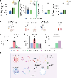
Fig. 5 |. Inhibition of endosomal escape detection has positive effects on mRNA expression and non-inflammatory LNPs abrogate ARDS.
a, In RAW 264.7 macrophages, TG pre-treatment increases luciferase mRNA expression by >2-fold. b, TG pre-treatment does not attenuate expression in the lung when TG and LNPs are administered intratracheally. c, TG pre-treatment improves mRNA expression in the liver and spleen by ~2.7 and ~2.4-fold, respectively, when TG and LNPs are injected intravenously. In all experiments, TG was administered 1 h before LNPs, and luminescence measurements were taken 6 h after LNP treatment. d, Timeline of treatments in nebulized-LPS ARDS model with LNPs containing mRNA encoding for the anti-inflammatory cytokine IL-10. e, IL-10 mRNA-loaded 4A3-SC8 LNPs and TG + IL-10 mRNA-loaded cKK-E12 LNPs induced upregulation of IL-10 by ~40-fold and ~60-fold, respectively, in the BAL fluid. f,g, IL-10 4A3-SC8 LNPs and TG + IL-10 cKK-E12 LNPs completely abrogated leukocyte infiltration (f) (BAL leukocyte count) and capillary leak into the alveolar space (g) (BAL protein levels). h, Summary schematic showing the two strategies for ameliorating LNP-induced inflammation while maintaining high mRNA expression, namely utilizing ESCRT-recruiting ionizable lipids and inhibiting galectins. Statistics: for ac, n = 3. For eg, n = 4. The data shown represent mean ± s.e.m. For ac, comparisons between groups were made using an unpaired two-tailed t-test with Welch’s correction. For all other graphs, comparisons between groups were made using one-way ANOVA with Tukey’s post hoc test. All graphics created in BioRender. Brenner, J. (2025) https://BioRender.com/b33cx8z.

Similar articles

References

    1. Kon E, Elia U & Peer D Principles for designing an optimal mRNA lipid nanoparticle vaccine. Curr. Opin. Biotechnol. 73, 329–336 (2022). - PMC - PubMed
    1. Verma M et al. The landscape for lipid-nanoparticle-based genomic medicines. Nat. Rev. Drug Discov. 22, 349–350 (2023). - PubMed
    1. Kiaie SH et al. Recent advances in mRNA-LNP therapeutics: immunological and pharmacological aspects. J. Nanobiotechnol. 20, 276 (2022). - PMC - PubMed
    1. Baden LR et al. Efficacy and safety of the mRNA-1273 SARS-CoV-2 vaccine. N. Engl. J. Med. 384, 403–416 (2021). - PMC - PubMed
    1. Polack FP et al. Safety and efficacy of the BNT162b2 mRNA Covid-19 vaccine. N. Engl. J. Med. 383, 2603–2615 (2020). - PMC - PubMed

Grants and funding

LinkOut - more resources