. 2025 Aug 11;15(1):29314.
doi: 10.1038/s41598-025-14956-3.

High-energy X-ray phase-contrast CT of an adult human chest phantom

Affiliations

High-energy X-ray phase-contrast CT of an adult human chest phantom

Jannis N Ahlers et al. Sci Rep. .

Abstract

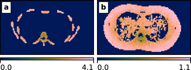
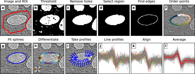
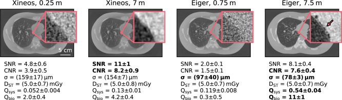
Propagation-based phase-contrast X-ray imaging is a promising technique for in vivo medical imaging, offering lower radiation doses than traditional attenuation-based imaging. Previous studies have focused on X-ray energies below 50keV for small-animal imaging and mammography. Here, we investigate the feasibility of high-energy propagation-based computed tomography for human adult-scale lung imaging at the Australian Synchrotron's Imaging and Medical Beamline. This facility is uniquely positioned for human lung imaging, offering a large field of view, high X-ray energies, and supporting clinical infrastructure. We imaged an anthropomorphic chest phantom (LungMan) between 50keV and 80keV across the range of possible sample-to-detector distances, with a photon-counting and an integrating detector. Strong phase-contrast fringes were observed with the photon-counting detector, even at high X-ray energies and a large pixel size relative to previous work, whereas the integrating detector with lower spatial resolution showed no clear phase effects. Measured X-ray phase-shifting properties of LungMan aligned well with reference soft tissue values, validating the phantom for phase-contrast studies. Imaging quality assessments suggest an optimal configuration at approximately 70keV and the longest available propagation distance of 7.5m, indicating potential benefit in positioning the patient in an upstream hutch. This study represents the first step towards clinical adult lung imaging at the Australian Synchrotron.

Keywords: Lung; Lungman; Phase-contrast; Propagation-based; X-ray.


Conflict of interest statement

Declarations. Competing interests: The authors declare no competing interests.

Figures

Fig. 1
Fig. 1
Experimental setup in hutch 3B of the Imaging and Medical Beamline (IMBL), directly downstream of hutch 3A. The CT stage can be moved along the beam path (shown in red). As Eiger is set back by 50 cm on the detector table, the propagation distances for Eiger datasets were always 50 cm more than for Xineos datasets. The inset images show (a) the tree insert, composed of a mediastinum with attached pulmonary vessels, and (b) the foam insert, while the inset diagram (c) shows the approximate size and position of the beam on LungMan.
Fig. 2
Fig. 2
Incident air kerma to local absorbed dose conversion coefficients for (a) cortical bone and (b) soft tissue at 70keV.
Fig. 3
Fig. 3
The fileswell algorithm for semi-automated line profiling. (a) The input image and region of interest. (b) Binary thresholding. (c) Hole filling. (d) Selection of the largest region within the field of view. (e) Edge detection using the Canny algorithm. (f) Ordering of the edge points using the travelling salesman algorithm. (g) Spline interpolation of the ordered edge points. (h) Differentiation of the spline to get the local gradients. (i) Taking line profiles, that fit within the ROI, across the interface. (j) All line profiles across the interface. (k) Aligning the line profiles. (l) Averaging to get the mean profile and the standard deviation of the points within that profile.
Fig. 4
Fig. 4
Comparing features in flat-field corrected and stitched projections of LungMan taken with Eiger and Xineos. In the central image, Eiger contains the tree insert. It is a composite image, with each quadrant showing a whole-lung stitched projection with a respective detector and propagation distance. In addition, we show a small region of interest from images taken of Eiger containing the foam insert (the approximate position of the ROI is shown on the composite image). While phase fringes and speckle patterns clearly develop in the Eiger images taken at the longer propagation distance, they cannot be seen with the Xineos detector. Note that the images are stitched from approximately 3 cm to 5 cm tall projections in which the flat-field illumination strongly varied. In addition, the Eiger projections were taken with an exposure time three times longer than the Xineos projections. Therefore, the dose cannot be directly compared between the different images in this figure; the figure is included to show the qualitative features that were present. The reader is referred to Fig. 5 for a dose-matched and quantitative comparison of the two detectors.
Fig. 5
Fig. 5
Comparison of phase-retrieved CT slices at the same mean absorbed dose of approximately 5mGy. All images were taken at 70keV. See Sect. "Imaging quality analysis" for definitions of the quality metrics. While the signal- and contrast-to-noise ratios are similar for Xineos and Eiger at the longest distances, the significantly better resolution means that imaging qualities are much higher using Eiger at 7.5m than for any other combination. The zoomed insets highlight the boundary between the mediastinum and a primary bronchus, which is filled with light foam for structural stability (intended to be transparent in conventional X-ray attenuation imaging). The small air bubbles at the edge of that foam can be clearly seen with Eiger at 7.5m (see arrow), but are not visible in the other images. Slices are shown at a greyscale of formula image to formula image, with insets set to the local min/max.
Fig. 6
Fig. 6
Measurement of the phase-shifting properties of LungMan. (a) In a central (high dose, low noise) slice of the dataset, a number of air–soft tissue interfaces are manually selected. (b) For each region of interest, the fringe between the two materials is profiled using the fileswell algorithm. (c) The resulting fringe profile is fit with a modified error function model (Eq. 5) to find formula image.
Fig. 7
Fig. 7
Results of the measurement of delta (formula image) values of the soft tissue equivalent material in LungMan at 50, 60, 70 and 80 keV. The results show excellent agreement with reference values for soft tissue. For context, reference values for cortical bone, which differ strongly from the measured values, are also shown. Error bars on the measurements show one standard deviation.
Fig. 8
Fig. 8
Results of imaging quality analysis. The metrics outlined in Sect. "Imaging quality analysis" are plotted against X-ray energy and propagation distance. The mean value at each combination is shown in the filled circle. As expected, larger propagation distances show increased SNR, but also reduced spatial resolution. Combining fluence-/dose-normalised SNR/CNR and resolution (Figs. 8c to 8e) suggests an optimal setup at 70keV and 7.5m.

References

    1. Snigirev, A., Snigireva, I., Kohn, V., Kuznetsov, S. & Schelokov, I. On the possibilities of x-ray phase contrast microimaging by coherent high-energy synchrotron radiation. Rev. Sci. Instruments66, 5486–5492. 10.1063/1.1146073 (1995).
    1. Cloetens, P., Barrett, R., Baruchel, J., Guigay, J.-P. & Schlenker, M. Phase objects in synchrotron radiation hard x-ray imaging. J. Phys. D: Appl. Phys.29, 133. 10.1088/0022-3727/29/1/023 (1996).
    1. Wilkins, S. W., Gureyev, T. E., Gao, D., Pogany, A. & Stevenson, A. W. Phase-contrast imaging using polychromatic hard X-rays. Nature384, 335–338. 10.1038/384335a0 (1996).
    1. Gureyev, T. E. et al. Refracting Röntgen’s rays: Propagation-based x-ray phase contrast for biomedical imaging. J. Appl. Phys.105, 10.1063/1.3115402 (2009).
    1. Paganin, D., Mayo, S. C., Gureyev, T. E., Miller, P. R. & Wilkins, S. W. Simultaneous phase and amplitude extraction from a single defocused image of a homogeneous object. J. Microsc.206, 33–40. 10.1046/j.1365-2818.2002.01010.x (2002). - PubMed