

Improvement in matching lesions in dual-view mammograms using a geometric model
- PMID: 40790177
- PMCID: PMC12337522
- DOI: 10.1186/s12880-025-01862-3
Improvement in matching lesions in dual-view mammograms using a geometric model
Abstract
Objectives: To evaluate the effectiveness of a geometric model (GM) as an adjunctive tool for radiologists to match lesions between craniocaudal (CC) and mediolateral (MLO) views.
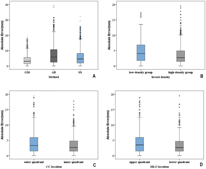
Methods: A retrospective study was conducted on 711 patients who underwent mammography from January 2016 to August 2018. Two senior radiologists used bounding boxes to delineate lesions as the reference standard, calculated the absolute error (the shortest distance from the lesion center to the predicted curve) of GM, and compared it with the annular band (AB) and straight strip (SS) methods. Four radiologists of varying seniority levels were tasked with localizing the corresponding lesion in MLO view using a bounding box, based on the given lesion in CC views, and recording reading time per case with or without GM assistance. The Dice coefficient was used to evaluate the overlap between the bounding box and the reference standard.
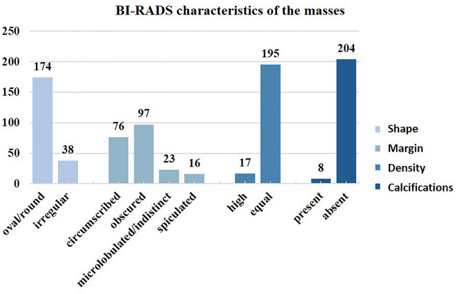
Results: Overall, 499 calcification and 212 mass pairs were evaluated. GM outperformed both AB and SS, yielding a median absolute error of 3.03 mm (IQR 1.45-5.55 mm) versus 5.78 mm (IQR 2.44-10.71 mm) for AB and 4.59 mm (IQR 1.91-8.19 mm) for SS (P < 0.001). With GM assistance, all four radiologists achieved improved Dice coefficients and reduced reading times (all P < 0.001). Stratified analysis by lesion conspicuity demonstrated that GM assistance significantly enhanced Dice coefficients for all radiologists in the low-conspicuity group and improved matching consistency for junior radiologists.
Conclusion: The geometric model holds substantial promise as a valuable tool to assist radiologists in more effectively localizing lesions in ipsilateral mammograms, thereby potentially enhancing diagnostic accuracy and efficiency.
Keywords: Breast; Craniocaudal; Image matching; Mammography; Mediolateral oblique.
© 2025. The Author(s).
Conflict of interest statement
Declarations. Ethics approval and consent to participate: In accordance with the Declaration of Helsinki, ethical approval was provided by the Ethics Committee of Nanfang Hospital of Southern Medical University, and the requirement for informed consent from patients was waived. Consent for publication: Not applicable. Competing interests: The authors declare no competing interests.
Figures


References
-
- Sung H, Ferlay J, Siegel RL, et al. Global cancer statistics 2020: GLOBOCAN estimates of incidence and mortality worldwide for 36 cancers in 185 countries. CA Cancer J Clin. 2021;71(3):209–49. - PubMed
-
- Amit G, Hashoul S, Kisilev P, Ophir B, Walach E, Zlotnick A. Automatic dual-view mass detection in full-field digital mammograms. In MICCAI. Springer, 2015: 44–52.
-
- Yan Y, Conze PH, Lamard M, Quellec G, Cochener B, Coatrieux G. Towards improved breast mass detection using dual-view mammogram matching. Med Image Anal. 2021;71:102083. - PubMed
-
- Velikova M, Samulski M, Lucas PJ, Karssemeijer N. Improved mammographic CAD performance using multi-view information: a bayesian network framework. Phys Med Biol. 2009;54(5):1131–47. - PubMed
-
- Lauby-Secretan B, Scoccianti C, Loomis D, et al. Breast-Cancer screening --Viewpoint of the IARC working group. N Engl J Med. 2015;372(24):2353–8. - PubMed
MeSH terms
Grants and funding
- 2019CR003/Clinical Research Program of Nanfang Hospital, Southern Medical University
- 2023B049/President's Fund of Nanfang Hospital, Southern Medical University
- 82171929/the National Natural Science Foundation of China
- 320.6750.2021-06-36/Wu Jieping Medical Foundation
- 2024A1111120015/Science and Technology Program of Guangdong Province, China
LinkOut - more resources
Full Text Sources
Medical
