Review
. 2025 Aug 11;32(1):75.
doi: 10.1186/s12929-025-01170-6.

Nuclear PKM2: a signal receiver, a gene programmer, and a metabolic modulator

Affiliations
Review

Nuclear PKM2: a signal receiver, a gene programmer, and a metabolic modulator

Tsan-Jan Chen et al. J Biomed Sci. .

Abstract

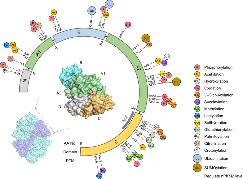
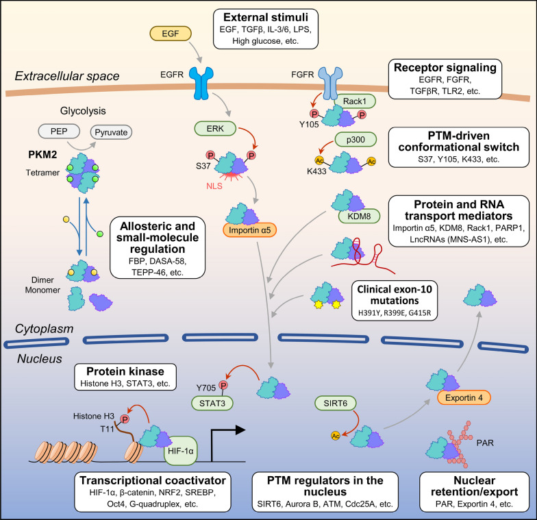
Pyruvate kinase M2 (PKM2) is a key enzyme involved in glycolysis, yet its role in cancer extends far beyond metabolic flux. Unlike its isoform PKM1, PKM2 exhibits unique regulatory properties due to alternative splicing and dynamic structural plasticity, enabling it to translocate into the nucleus. Once nuclear, PKM2 functions as a signal receiver, gene programmer, and metabolic modulator by acting as a co-transcriptional activator and protein kinase. In this capacity, nPKM2 (nuclear PKM2) orchestrates the transcription of genes involved in glycolysis, lipogenesis, redox homeostasis, and cell cycle progression, thereby reinforcing the Warburg effect and promoting tumor growth, metastasis, and resistance to stress. In this regard, nPKM2 can be considered as the oncogenic component of PKM2. This review consolidates current knowledge on the structural basis of PKM2 assembly and the post-translational modifications that govern its oligomeric state and nuclear import. We also explore emerging therapeutic strategies aimed at targeting nPKM2, including small-molecule modulators that stabilize its cytosolic tetrameric form or disrupt its nuclear functions. Ultimately, the multifaceted roles of nuclear PKM2 underscore its significance as a critical oncoprotein and a promising target for precision cancer therapy.

Keywords: Cancer metabolism; Gene programmer; Metabolic modulator; Nuclear PKM2; Nuclear translocation; Oncogenic signaling; Post-translational modification; Signal receiver.


Conflict of interest statement

Declarations. Ethics approval and consent to participate: Not applicable. Consent for publication: Not applicable. Competing interests: The authors declare no competing interests.

Figures

Fig. 1
Fig. 1
Structural organization and allosteric regulation of PKM2. A The PKM gene undergoes mutually exclusive alternative splicing, yielding two isoforms, PKM1 (exon 9) and PKM2 (exon 10). The sequence alignment below highlights the 56–amino acid difference encoded by exon 10 that underlies unique regulatory properties. B Linear domain map of PKM2: the N domain (residues 1–43), the A domain (A1: residues 44–116 and A2: residues 219–389), the B domain (residues 117–218), and the C domain (residues 390–531). Shaded ticks mark the exon 10 region, nuclear localization signal (NLS) with R399/R400 marked with a black stick, active-site, amino-acid, fructose-1,6-bisphosphate (FBP), and small-molecule activator (DASA-58/TEPP-46) binding sites, as well as the AA and CC interfaces. C Surface model of one PKM2 subunit colored by domain (N, gray; A1/A2, green; B, cyan; C, ochre). Key functional sites are labeled: active site (PEP·ADP·K+·Mg2+), amino-acid site (Ser/Phe), FBP site, and activator site (DASA-58/TEPP-46). Dashed lines indicate the AA and CC interfaces that mediate tetramer assembly. D Conformational equilibrium of PKM2: the high-activity R-state tetramer (left) and the low-activity T-state tetramer (center) interconvert with dissociated dimer/monomer forms (right). Endogenous metabolites (serine, SAICAR, butyrate) and synthetic activators (DASA-58, TEPP-46) modulate this equilibrium toward the R-state, thereby enhancing catalytic output and blocking the structural transitions required for PKM2’s nuclear import and transcriptional functions. The structural models are illustrated based on PDB models 3SRD and 4FXJ
Fig. 2
Fig. 2
Post-translational modification landscape of PKM2. The circular schematic depicts PKM2’s primary structure (residues 1–531), color-coded by domain: N (gray), A1 (green), B (cyan), A2 (green), and C (ochre), with the nuclear localization signal (NLS) indicated in dark gray. Around the periphery, icons mark the positions and types of known PTMs: phosphorylation (P), acetylation (Ac), hydroxylation (OH), oxidation (Ox), O-GlcNAcylation (Gly), succinylation (Suc), methylation (Me), lactylation (Lac), sulfhydration (SSH), glutathionylation (SSG), palmitoylation (Pal), citrullination (Cit), crotonylation (Cr), ubiquitination (Ub), and SUMOylation (SU). Red connectors highlight those PTMs experimentally shown to regulate PKM2 nuclear import. The inset shows a PKM2 tetramer (surface view), with one subunit zoomed in to illustrate how PTMs cluster at key interdomain interfaces and regulatory sites
Fig. 3
Fig. 3
Sporadic PKM2 mutations in the exon 10 region and their association with oligomeric assembly. This schematic illustrates the domain architecture of PKM2 (residues 1–531), highlighting the core pyruvate kinase (PK) domain (green) and the PK_C domain (red). Each mutation from TCGA data, with several mutations clustering within the exon 10 region that is clinically relevant for allosteric regulation and oligomerization (https://www.cbioportal.org/)
Fig. 4
Fig. 4
Integrated model of PKM2 nuclear translocation and nuclear functions. The schematic diagram illustrates how PKM2 shuttles between the cytosol and nucleus in response to metabolic cues, post-translational modifications (PTMs), and protein- or RNA-mediated interactions

References

    1. Anastasakis DG, Apostolidi M, Garman KA, Polash AH, Umar MI, Meng Q, Scutenaire J, Jarvis JE, Wang X, Haase AD, Brownell I, Rinehart J, Hafner M. Nuclear PKM2 binds pre-mRNA at folded G-quadruplexes and reveals their gene regulatory role. Mol Cell. 2024;84(19):3775–89. - PMC - PubMed
    1. Anastasiou D, Poulogiannis G, Asara JM, Boxer MB, Jiang JK, Shen M, Bellinger G, Sasaki AT, Locasale JW, Auld DS, Thomas CJ, Vander Heiden MG, Cantley LC. Inhibition of pyruvate kinase M2 by reactive oxygen species contributes to cellular antioxidant responses. Science. 2011;334(6060):1278–83. - PMC - PubMed
    1. Anastasiou D, Yu Y, Israelsen WJ, Jiang JK, Boxer MB, Hong BS, Tempel W, Dimov S, Shen M, Jha A, Yang H, Mattaini KR, Metallo CM, Fiske BP, Courtney KD, Malstrom S, Khan TM, Kung C, Skoumbourdis AP, Veith H, Southall N, Walsh MJ, Brimacombe KR, Leister W, Lunt SY, Johnson ZR, Yen KE, Kunii K, Davidson SM, Christofk HR, Austin CP, Inglese J, Harris MH, Asara JM, Stephanopoulos G, Salituro FG, Jin S, Dang L, Auld DS, Park HW, Cantley LC, Thomas CJ, Vander Heiden MG. Pyruvate kinase M2 activators promote tetramer formation and suppress tumorigenesis. Nat Chem Biol. 2012;8(10):839–47. - PMC - PubMed
    1. Angiari S, Runtsch MC, Sutton CE, Palsson-McDermott EM, Kelly B, Rana N, Kane H, Papadopoulou G, Pearce EL, Mills KHG, O’Neill LAJ. Pharmacological activation of pyruvate kinase M2 inhibits CD4(+) T cell pathogenicity and suppresses autoimmunity. Cell Metab. 2020;31(2):391–405. - PMC - PubMed
    1. Anitha M, Kaur G, Baquer NZ, Bamezai R. Dominant negative effect of novel mutations in pyruvate kinase-M2. DNA Cell Biol. 2004;23(7):442–9. - PubMed