. 2025 Aug 11;15(1):29378.
doi: 10.1038/s41598-025-10980-5.

Unveiling the hidden culturable endophytic fungal diversity in aerial vegetative parts of Wrightia tinctoria (Roxb.) R.Br. of southern Aravalli hills

Affiliations

Unveiling the hidden culturable endophytic fungal diversity in aerial vegetative parts of Wrightia tinctoria (Roxb.) R.Br. of southern Aravalli hills

Garima Yadav et al. Sci Rep. .

Abstract

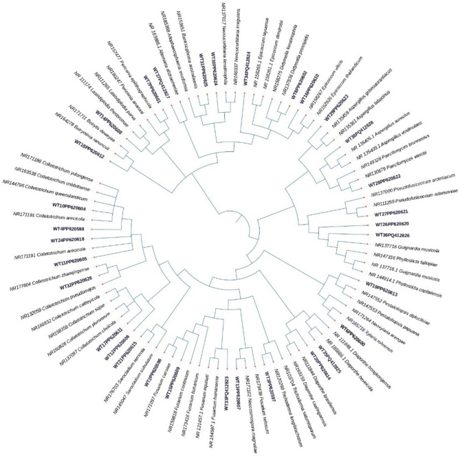
Endophytic fungi significantly influence plant health, growth, and ecological interactions, yet comprehensive insights into their diversity within medicinal plants remain limited. This study provides the first comprehensive analysis of alpha and beta diversity of fungal endophytic communities residing within Wrightia tinctoria (Roxb.) R.Br., examining variations across different plant parts (leaf, stem, and bark), seasons, and geographic locations. A total of 3929 fungal isolates representing 32 morphotypes, primarily from the phylum Ascomycota, were isolated from 6075 tissue segments. Notably, Fusarium cassiae, Neocosmospora magnoliae, Xylaria rohrensis, and Pestalotiopsis papuana were globally reported as endophytes for the first time. Colonization frequency varied significantly with maximum colonization observed at location 1 (80.88%), specifically in leaf tissues (84.64%) and during the monsoon season (80.91%). Analyses of alpha and beta diversity revealed marked variations in locations, plant parts, and seasons. Beta diversity analyses further illustrated both unique and overlapping fungal communities across different conditions, supported by non-metric multidimensional scaling and hierarchical clustering based on Bray-Curtis dissimilarity. Principal component analysis indicated that the first two components explained 40.4% of the observed diversity variations, primarily influenced by location, plant part, and seasonal dynamics. The study concludes that fungal endophytic diversity within W. tinctoria is significantly structured by ecological factors such as plant tissue type, seasonal variation, and geographic location, emphasizing the complexity and specificity of plant-endophyte interactions.

Keywords: Distribution; Diversity analysis; Fungal endophytes; Geographical locations; Phylogenetic analysis; Seasonal variations.


Conflict of interest statement

Declarations. Competing interests: The authors declare no competing interests.

Figures

Fig. 1
Fig. 1
Evolutionary tree constructed using maximum likelihood method using internal transcribed spacer (ITS)–rDNA gene sequences of fungal endophytes isolated from Wrightia tinctoria and their closest sequences procured from GenBank.
Fig. 2
Fig. 2
Bar chart displaying significant variation at p ≤ 0.0001 in colonization frequency with respect to different seasons (a), locations (b) and tissue types (c).
Fig. 3
Fig. 3
Heatmaps demonstrating the relative abundance of isolated fungal endophyte across different seasons (a), locations (b), and plant parts (c) where colour gradient represents relative abundance with orange indicating higher abundance and teal lower abundance, and dendrogram showing clustering of the morphotypes on the basis of similarities in their abundance pattern across different seasons (a), locations (b), and plant parts (c).
Fig. 4
Fig. 4
The non-metric multidimensional scaling (nMDS) plot represents the variation in fungal community composition based on plant parts, seasons, and locations. Axes (nMDS1 and nMDS2) denote two dimensions of endophytic fungal communities, where the lesser distance between the points, the more similar the fungal endophytic composition. On the other hand, vectors represent the impact of different variables on the shaping of fungal community structure. This plot signifies the relationships among endophytic fungal communities from different seasons, plant parts, and locations.
Fig. 5
Fig. 5
Pairwise comparison of fungal endophytic communities based on season, location, and plant part. Comparison of fungal communities across different seasons (a), locations (b), and plant parts (c), where the scatter plots reflect the distribution of fungal endophytes in different seasons, locations and plant parts, with confidence ellipses representing the 95% confidence interval for each variable.
Fig. 6
Fig. 6
The PCA analysis represents the distribution of several fungal endophytes across the first two principal components, PC1 (24.0%) and PC2 (16.4%). Each point represents a fungal isolate, with symbols denoting plant tissue types and colors indicating collection locations. Symbol shapes: ★ = Bark, ◆ = Stem, ● = Leaf. Colors represent locations: Green = Loc 1, Purple = Loc 2, Red = Loc 3. The fungal isolates are scattered with respect to their variance contributions across the PCA axes, where arrows demonstrate the loading vectors for the corresponding fungal endophyte, reflecting their relative significance in contributing to the variation along PC1 and PC2.
Fig. 7
Fig. 7
Boxplot illustrating alpha diversity variation across different seasons, locations, and plant parts. Shannon, Simpson’s and Fisher’s_alpha indices had no significant variances (p > 0.05) across all seasons (a, b, c). Shannon, Simpson’s and Fisher’s_alpha indices had significant variances (p ≤ 0.0001) across all sampling locations (d, e, f). Shannon, Simpson’s and Fisher’s_alpha indices had significant variances at (p > 0.01) and (p > 0.05) among different plant parts (g, h, i).

References

    1. Ghosh, A. et al. Crystal structure and DFT calculations of 3, 4-seco-lup-20 (29)-en-3-oic acid isolated from Wrightia tinctoria: Stacking of supramolecular dimers in the crystal lattice. J. Mol. Struct.980, 7–12 (2010).
    1. Chen, Y.-J., Chen, H.-J. & Chung, W.-H. Endophytic fungal diversity in Cirsium kawakamii from Taiwan. J. Fungi9, 1076 (2023). - PMC - PubMed
    1. Yadav, G. & Meena, M. Bioprospecting of endophytes in medicinal plants of Thar Desert: An attractive resource for biopharmaceuticals. Biotechnol. Rep.30, e00629 (2021). - PMC - PubMed
    1. White, J. F. et al. Evidence for widespread microbivory of endophytic bacteria in roots of vascular plants through oxidative degradation in root cell periplasmic spaces. In PGPR Amelioration in Sustainable Agriculture 167–193 (Elsevier, 2019).
    1. Trivedi, P., Leach, J. E., Tringe, S. G., Sa, T. & Singh, B. K. Plant–microbiome interactions: From community assembly to plant health. Nat. Rev. Microbiol.18, 607–621 (2020). - PubMed