. 2025 Aug;21(8):e70561.
doi: 10.1002/alz.70561.

Glymphatic activity in behavioral variant frontotemporal dementia: Link with vascular function and astrocytic activation

Affiliations

Glymphatic activity in behavioral variant frontotemporal dementia: Link with vascular function and astrocytic activation

Min Chu et al. Alzheimers Dement. 2025 Aug.

Abstract

Introduction: Glymphatic activity, vascular function, and astrocytic activation play pivotal roles in the pathophysiology of frontotemporal dementia (FTD). However, their interrelationships and combined impact on clinical features remain unclear.

Methods: Glymphatic activity was measured by diffusion tensor image analysis along the perivascular space (DTI-ALPS) in patients with behavioral variant FTD (bvFTD, n = 61) and normal controls (NC, n = 61).

Results: Glymphatic activity in bvFTD correlated strongly with vascular dysfunction and astrocytic activation, and was associated with brain changes and clinical severity. Astrocytic activation mediates the association between vascular and glymphatic function. Astrocytic activation mediates the association between glymphatic function and clinical scales, whereas glymphatic function mediates the association between vascular function and clinical scales. The interaction effect was found between vascular function and glymphatic activity on white matter hyperintensity.

Discussion: Our findings reveal a complex interrelationship among glymphatic, vascular, and inflammatory mechanisms in FTD, highlighting their collective impact on disease severity. Highlight Vascular function, astrocytic activation, and glymphatic activity correlate with each other in frontotemporal dementia. Glymphatic activity mediates the association between vascular function and cognition. Astrocytic activation mediates the association between glymphatic function and cognition. Vascular and glymphatic function synergy drives white matter hyperintensity.

Keywords: astrocytic activation; frontotemporal dementia; glymphatic system; vascular function.


Conflict of interest statement

The authors declare no conflicts of interest. Any author disclosures are available in the Supporting Information.

Figures
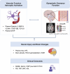
FIGURE 1
FIGURE 1
Flowchart of the study. Patients with bvFTD and normal controls are enrolled and complete plasma biomarkers testing, clinical scales, multimodal MRI, and PET imaging. We tested vascular function biomarkers, including plasma galectin‐3, MMP‐9, VCAM1, PDGF‐BB, and TGF‐β1; astrocytic activation biomarker, including plasma GFAP; glymphatic clearance dysfunction biomarkers, including DTI‐ALPS; neural injury biomarkers, including plasma t‐tau and NfL; brain changes biomarker, including brain atrophy, FDG hypometabolism, and WMH; clinical outcomes, including MMSE, MoCA, CDR plus NACC FTLD, FBI, TMT, BNT, and ADL scales. We aimed to elucidate the interplay among glymphatic, vascular, and inflammatory mechanisms and evaluate their contributions to neural injury, brain changes, and clinical outcomes. The figure was created with BioRender.com. bvFTD, behavioral variant frontotemporal dementia; MMP, matrix metalloproteinase; VCAM1, vascular cell adhesion molecule‐1; GFAP, glial fibrillary acidic protein; NfL, neurofilament light chain; PDGF‐BB, platelet‐derived growth factor‐BB; t‐tau, total tau; TGF‐β1, transforming growth factor‐beta 1; DTI‐ALPS, diffusion tensor image analysis along the perivascular space; FDG, fluorodeoxyglucose (uptake); WMH, white matter hyperintensity; MMSE, Mini‐Mental State Examination; MoCA, Montreal Cognitive Assessment; CDR plus NACC FTLD‐SB, the Sum of the Boxes scores of Clinical Dementia Rating plus The National Alzheimer's Coordinating Center Frontotemporal Lobar Degeneration; TMT, Trail Making Test; FBI, Frontal Behavior Inventory; BNT, Boston Naming Test; ADL, Activities of Daily Living scale.
FIGURE 2
FIGURE 2
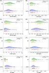
Decreased DTI‐ALPS was observed in the bvFTD group. Significantly reduced anterior ALPS (aALPS,A‐1), middle ALPS (mALPS, A‐2), and global ALPS (gALPS, B‐2) values were observed in the bvFTD group (Model 3 covariate: adjusted for sex, age, education, and corresponding FA and MD), no difference was found in posterior ALPS (B‐1), indicating impairment of the glymphatic system. bvFTD, behavioral variant frontotemporal dementia; DTI‐ALPS, diffusion tensor image analysis along the perivascular space.
FIGURE 3
FIGURE 3
Alterations of plasma vascular, astrocytic activation, and neural injury biomarkers in the FTD group. The group comparison (Model 3: adjusted for sex, age, and education) shows plasma levels of MMP‐9 (B), VCAM1 (E), GFAP (F), and NfL (G) were significantly elevated. In contrast, PDGF‐BB (C) and TGF‐β1 (D) levels were decreased in the FTD group. No significant changes were observed in galectin‐3 (A) and t‐tau (H). bvFTD, behavioral variant frontotemporal dementia; MMP, matrix metalloproteinase; VCAM1, vascular cell adhesion molecule‐1; GFAP, glial fibrillary acidic protein; NfL, neurofilament light chain; PDGF‐BB, platelet‐derived growth factor‐BB; t‐tau, total tau; TGF‐β1, transforming growth factor‐beta 1.
FIGURE 4
FIGURE 4
Partial correlation analysis between biomarkers of glymphatic activity, vascular function, astrocytic activation, and neural injury. Positive correlations are represented in red, and negative correlations are represented in blue. Significance is displayed within the corresponding boxes and denoted by asterisks: *p < 0.05, **p < 0.01, ***p < 0.001, ****p < 0.0001. Glymphatic function is represented by aALPS, mALPS, pALPS, and gALPS; vascular function includes plasma galectin‐3, MMP‐9, PDGF‐BB, VCAM1, and TGF‐β1; GFAP indicates astrocytic activation; and plasma NfL and t‐tau reflect neural injury. Significant correlations before FDR correction (Model 3): aALPS and mALPS, aALPS and pALPS, aALPS and gALPS, aALPS and VCAM1, aALPS and GFAP, mALPS and pALPS, mALPS and gALPS, pALPS and gALPS, gALPS and VCAM1, gALPS and GFAP, PDGF‐BB and TGF‐β1, VCAM1 and GFAP, VCAM1 and t‐tau, GFAP and t‐tau. bvFTD, behavioral variant frontotemporal dementia; DTI‐ALPS, diffusion tensor image analysis along the perivascular space; a, anterior; m, middle; p, posterior; g, global; MMP, matrix metalloproteinase; VCAM1, vascular cell adhesion molecule‐1; PDGF‐BB, platelet‐derived growth factor‐BB; t‐tau, total tau; TGF‐β1, transforming growth factor‐beta 1; GFAP, glial fibrillary acidic protein; NfL, neurofilament light chain.
FIGURE 5
FIGURE 5
Relationship between glymphatic activity, vascular function, astrocytic activation, brain changes, and clinical outcomes. This figure shows the mediation and interaction results of Model 3. (A) Plasma GFAP acts as a mediator in the relationship between plasma VCAM1 and aALPS. Conversely, plasma VCAM1 did not serve as a mediator between plasma GFAP and aALPS. (B) Plasma GFAP mediates the relationship between aALPS and clinical scales, whereas aALPS mediates the relationship between plasma VCAM1 and clinical scales. (C)The interaction effect of plasma VCAM1 and aALPS on white matter hyperintensities. aALPS, anterior diffusion tensor image analysis along the perivascular space; CDR plus NACC FTLD‐SB, the Sum of Boxes scores of the 6 domains of the CDR plus the behavior/comportment/personality and language domains; MoCA, Montreal Cognitive Assessment; VCAM1, vascular cell adhesion molecule‐1; GFAP, glial fibrillary acidic protein; WMH, white matter hyperintensity; PVWMH, periventricular white matter hyperintensity; DWMH, deep white matter hyperintensity.

References

    1. Bang J, Spina S, Miller BL. Frontotemporal dementia. Lancet. 2015;386:1672‐1682. - PMC - PubMed
    1. Nedergaard M, Goldman SA. Glymphatic failure as a final common pathway to dementia. Science. 2020;370:50‐56. - PMC - PubMed
    1. Ringstad G, Vatnehol SAS, Eide PK. Glymphatic MRI in idiopathic normal pressure hydrocephalus. Brain. 2017;140:2691‐2705. - PMC - PubMed
    1. Jiang D, Liu L, Kong Y, et al. Regional glymphatic abnormality in behavioral variant frontotemporal dementia. Ann Neurol. 2023;94:442‐456. - PMC - PubMed
    1. Xiao D, Li J, Ren Z, et al. Association of cortical morphology, white matter hyperintensity, and glymphatic function in frontotemporal dementia variants. Alzheimers Dement. 2024;20:6045‐6059. - PMC - PubMed

LinkOut - more resources