[Preprint]. 2025 Jul 18:2025.07.15.664798.
doi: 10.1101/2025.07.15.664798.

Huntington's disease LIG1 modifier variant increases ligase fidelity and suppresses somatic CAG repeat expansion

Affiliations

Huntington's disease LIG1 modifier variant increases ligase fidelity and suppresses somatic CAG repeat expansion

Eunhye Lee et al. bioRxiv. .

Abstract

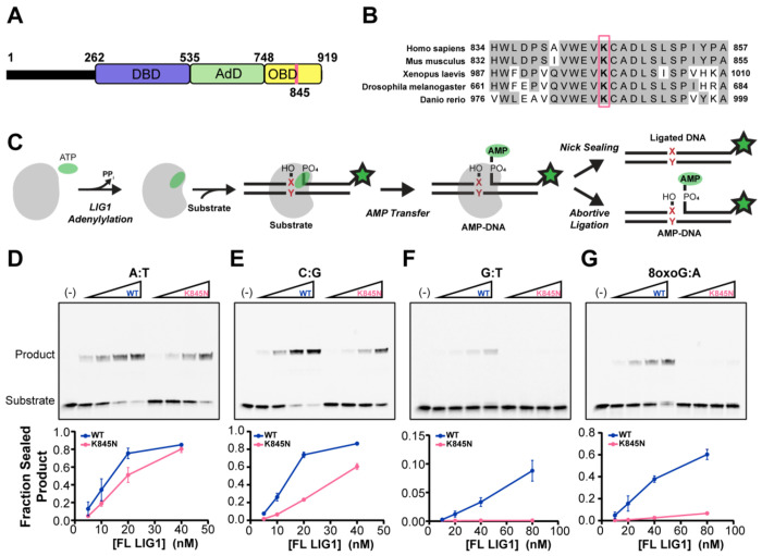
Huntington's disease (HD) is a fatal neurodegenerative disorder caused by inheriting an expanded CAG repeat tract in the huntingtin gene (HTT) that further expands in somatic cells over an individual's lifetime. Genome-wide association studies have provided critical insight into factors that modify the course of disease. These include DNA repair genes that alter the rate of somatic expansion and other genes that do not appear to directly influence this process. One modifier gene is DNA ligase 1 (LIG1), in which a variant specifying a lysine to asparagine substitution (K845N) is associated with a profound (7-8 year) delay in the onset of motor signs. Here, we have taken a multifaceted approach to gain insight into the protective nature of this variant in HD. We demonstrate using in vitro ligase assays and enzyme kinetics that K845N enhances discrimination towards mismatched substrates and increases repair fidelity. Consistent with increased ligation fidelity, K845N confers protection against oxidative stress in cell-based assays. Finally, we demonstrate that the mouse LIG1 K843N orthologue suppresses somatic CAG expansion in HD knock-in mice. Overall, our data provide evidence that altered LIG1 function due to the K845N substitution may contribute to HD clinical delay by slowing somatic expansion in the brain and protecting the genome globally against damage. Significantly, our results provide a mechanistic foundation for considering DNA ligase fidelity as a therapeutic target in HD and potentially in other trinucleotide repeat disorders.

Keywords: Biological Sciences; DNA damage; DNA ligase 1; Genetics; Huntington’s disease; repair fidelity; somatic repeat expansion.


Figures

Figure 1.
Figure 1.. LIG1 K845N shows reduced ligase activity towards mismatched substrates.
(A) Domain architecture of LIG1. The DNA binding domain (DBD), adenylylation domain (AdD), and OB-fold domain (OBD) comprise the catalytic core. (B) Multiple sequence alignment of LIG1 orthologs by Clustal Omega. The position of K845 is indicated by the red box. (C) Illustration of LIG1 reaction. LIG1 is first adenylylated by ATP at K568, forming a LIG1-AMP intermediate. The second step, adenyl transfer, involves the transfer of AMP from LIG1 to the 5’ phosphate end of the DNA nick, resulting in a DNA-AMP intermediate. Finally, in the nick sealing step, the 3’ hydroxyl group at the DNA nick attacks the 5’ phosphodiester bond, displacing AMP and sealing the DNA backbone. LIG1 may also release premature AMP-DNA intermediates, known as abortive ligation. Our in vitro ligation reaction uses nicked substrate generated by annealing three oligonucleotides, one of which is labeled with FAM (green star), allowing substrate, aborted DNA-AMP and ligated product to be distinguished. X:Y = canonical or mismatched base pair. (D-G) Representative gels and quantified fraction of ligation product showing the ligase activity of full-length LIG1 WT and K845N in the context of nucleotide pairs at the 3’ nick position. A:T (D), C:G (E), G:T (F), 8-oxoG:A (G) containing nicked 34mer DNA (300 nM) was incubated with increasing concentrations of LIG1 proteins for 5 min at 37 °C. Reactions were performed with 1 mM ATP, 10 mM MgCl2, 50 mM MOPS pH 7.5 and 150 mM NaCl. Reactants were analyzed on 15 % TBE-urea polyacrylamide mini gels (note that in these gels substrate and AMP-DNA are not resolved). The line graphs show the quantification of the fraction of ligated product from three independent experiments (Mean ± SD).
Figure 2.
Figure 2.. K845N is a higher fidelity, but lower activity LIG1 variant as determined by Michaelis-Menten kinetics.
(A) Representative denaturing PAGE showing in vitro ligation reaction with WT or K845N Δ232 LIG1 (1 nM) with C:G or 8-oxoG:A containing nicked DNA (500 nM), resolving ligated product, aborted product and substrate. Reactions were performed at 37 °C with 1 mM ATP, 2 mM MgCl2 (1.0 mM free Mg2). Time courses for ligation of 1000 nM C:G (B) and 8-oxoG:A (C) 28mer substrate were plotted as number of turnovers with 1 and 5 nM LIG1 WT and 1.5 and 10 nM LIG1 K845N, respectively. Michaelis-Menten dependence for C:G (D) and 8-oxoG:A (E) 28mer nicked DNA was determined with 0.2 mM ATP and 1.0 mM free Mg2+. (F) The fraction abortive ligation determined from initial rates of ligation. The catalytic efficiency for ligation (G) and the discrimination against 8-oxoG:A (H) were calculated from the kinetic parameters in panels D and E. Data reported are mean ± SD (N≥3) and are summarized in SI Appendix, Table S2.
Figure 3.
Figure 3.. Cells harboring the LIG1 K845N variant exhibit increased viability and decreased DNA mutation under conditions that induce DNA damage.
(A) HEK 293 cells (EV: empty vector-transfected or expressing LIG1 wild type (WT) or LIG1 K845N (K845N)) were treated with 8 μM menadione for 4 hours. Cells were washed in fresh medium and further incubated for the indicated times. Cellular viability was measured by CellTiter-Glo (CTG). Bar graph shows mean of 3 technical replicates ± standard deviation. Statistical significance was determined from 2-way ANOVA with Tukey’s multiple comparison test (****p < 0.0001, **p < 0.01, *p < 0.05). (B-C) HEK 293 cells (WT or K845N) were treated with menadione for 4 hours at the indicated concentrations and washed in fresh medium. After 48 hours recovery, the cells were harvested. (B) A portion of the harvested cells was used to measure cell viability by CTG. Bar graph shows mean % viability relative to baseline (0% menadione) of 2 technical replicates ± standard deviation (C) DNA was extracted from the remaining cells and mutation frequencies were determined by duplex sequencing. Bar graph shows mean % mutation frequency (MF) relative to baseline (0% menadione) of 2 technical replicates ± standard deviation. Statistical significance in B and C was determined from two-way ANOVA with Tukey’s multiple comparison test (*p < 0.05, ** p < 0.01, ***p < 0.001). (D) LCLs derived from HD patients (E) were treated with menadione for 4 hours at the indicated concentrations. LCLs were either homozygous for the LIG1 rs145821638 (GRCh38 - Chr19:48117686) reference C-allele (LIG1+/+) or heterozygous for the 19AM3 modifier variant A-allele (LIG1K845N/+). Cells were washed in fresh medium and incubated for 24 hours. Viability of each LCL was determined using CTG from the mean value of three biological replicates. Bar graph shows mean % viability relative to baseline (0% menadione) ± standard deviation (n = 4 LIG1+/+ independent LCLs, n = 3 independent LIG1K845N/+ LCLs). Statistical significance indicated in figure was determined by two-way ANOVA with Tukey’s multiple comparison test.
Figure 4.
Figure 4.. The orthologous K843N mutation suppresses somatic CAG expansion in mice.
(A) CRISPR-Cas9-mediated generation of the Lig1K843N knock-in allele. Top: The sequence is part of mouse Lig1 exon 26 (canonical transcript ENSMUST00000177588.10) with amino acid translation showing lysine (K) at amino acid 843 (orthologous to human K845). The targeting single guide RNA (sgRNA) and protospacer adjacent motif (PAM) are shown. A 150 nt single stranded oligonucleotide (not shown – see Materials and Methods) was used for homology-directed repair to mutate G>A changing K843 to asparagine (N) (pink box). This also introduced a silent C>T change within the PAM (green box). Bottom: Sanger sequence from a Lig1 wild-type mouse, and heterozygous and homozygous K843N knock-in mice. (B) Somatic CAG expansion indices (Min to Max box-whisker plots) from striatum and liver of HttQ111/+ mice with different Lig1 genotypes. 3 mo: Lig1+/+ N=2, Lig1K843N/+ N=2, Lig1K843N/K843N N=8. 6 mo: Lig1+/+ N=4, Lig1K843N/+ N=9, Lig1K843N/K843N N=7. 10 mo: Lig1+/+ N=9, Lig1K843N/+ N=9, Lig1K843N/K843N N=10. **p < 0.01; *** p < 0.001, ****p < 0.0001 (One way ANOVA, comparing all genotypes for each age and tissue, with Tukey’s multiple comparison test).
Figure 5.
Figure 5.. Schematic illustrating increased fidelity of ligation due to the K845N substitution
Top panel: Ligation of canonical base-paired substrate (black X:Y pair). (i) LIG1 WT produces mostly fully ligated DNA product (thick arrow and black depiction), with a small amount of aborted AMP-DNA (thin arrow and gray depiction). (ii) LIG1 K845N does not have a large impact on the proportions of ligated DNA and AMP-DNA. Bottom panel: Ligation of mismatched substrate (blue X:Y). (iii) LIG1 WT produces both fully ligated DNA product and aborted AMP-DNA (intermediate arrows). (iv) In contrast, LIG1 K845N produces less ligated product (thin arrow and gray depiction) and releases more aborted AMP-DNA (thick arrow and black depiction).

References

    1. Harper P. S., Huntington’s disease: a clinical, genetic and molecular model for polyglutamine repeat disorders. Philos Trans R Soc Lond B Biol Sci 354, 957–961 (1999). - PMC - PubMed
    1. The Huntington’s Disease Collaborative Research Group., A novel gene containing a trinucleotide repeat that is expanded and unstable on Huntington’s disease chromosomes. The Huntington’s Disease Collaborative Research Group. Cell 72, 971–983 (1993). - PubMed
    1. Swami M. et al. Somatic expansion of the Huntington’s disease CAG repeat in the brain is associated with an earlier age of disease onset. Hum Mol Genet 18, 3039–3047 (2009). - PMC - PubMed
    1. Kennedy L. et al. Dramatic tissue-specific mutation length increases are an early molecular event in Huntington disease pathogenesis. Hum Mol Genet 12, 3359–3367 (2003). - PubMed
    1. Mouro Pinto R. et al. Patterns of CAG repeat instability in the central nervous system and periphery in Huntington’s disease and in spinocerebellar ataxia type 1. Hum Mol Genet 29, 2551–2567 (2020). - PMC - PubMed

Publication types