[Preprint]. 2025 Jul 18:2025.07.17.662885.
doi: 10.1101/2025.07.17.662885.

An ancient evolutionary origin for IL-1 cytokines as mediators of immunity

Affiliations

An ancient evolutionary origin for IL-1 cytokines as mediators of immunity

Francisco Fontenla-Iglesias et al. bioRxiv. .

Abstract

Inflammation is a hallmark of immune responses. Its mechanistic underpinnings in mammals are well-defined: pro-inflammatory cytokines of the interleukin 1 (IL-1) superfamily establish and support microenvironments that promote immune cell activities. Despite a growing number of reports on inflammatory processes and components of the IL-1 signaling axis in several invertebrate lineages, orthologs of these central cytokines have not been detected outside of the jawed vertebrates. Here, protein structure prediction algorithms were applied to identify genes encoding a family of IL-1 proteins with homologs throughout Eumetazoa (termed "IL-1anc" to reflect their ancestral evolutionary origin). Using Petromyzon marinus (sea lamprey) and Strongylocentrotus purpuratus (purple sea urchin) as model systems, we demonstrate that IL-1anc proteins share important features with mammalian IL-1α/IL-1β including expression patterns, protein localization, and processing. Together, our data indicate that the IL-1 superfamily and associated circuitry represent a foundational module of animal immunity that far predates the jawed vertebrates.

Keywords: evolution; immune response; inflammation; interleukin-1; invertebrates.


Figures

Figure 1.
Figure 1.. IL-1 family protein structures from divergent Metazoa.
Comparison of the crystal structures of (A) human IL-1β (1ITB) with computationally determined structures of the IL-1 trefoil domain from (B) Petromyzon marinus (sea lamprey) PmarIL-1.3, (C) PmarIL-1.1, (D) PmarIL-1.2, (E) Strongylocentrotus purpuratus (purple sea urchin) SpurIL-1.2, (F) the cephalochordate Branchiostoma floridae BfloIL-1, (G) an arthropod, the crustacean Procambarus clarkii PclaIL-1.2, (H) a mollusk, the bivalve scallop Pecten maximus PmaxIL-1.1, and (I) a cnidarian, the anthozoan Nematostella vectensis NvecIL-1. All structures are aligned to the orientation of human IL-1β in (A). For (A, B, and E) the 12 β-strands are labeled with numbers and a small conserved α-helix region with the letter A, and they are shown from two orientations rotated 90 degrees around the vertical axis. (J) The predicted structure of the complete chain of Petromyzon marinus PmarIL-1.2 with the PDZ domain (blue) and the IL-1 trefoil domain (green) is shown from two orientations. The N-terminal region is shown in white, and the linker between the PDZ and IL-1 trefoil is shown in orange.
Figure 2.
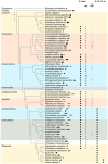
Figure 2.. IL-1anc homologs are present throughout metazoan lineages.
The distribution of IL-1 superfamily members is shown across Metazoa with representative species from major lineages. Filled circles indicate that at least one homolog is present in the respective lineage, with IL-1α and IL-1β being representative of the classical IL-1s whereas IL-1anc refers to the more widespread group described here. The numbers of predicted IL-1anc proteins with or without an N-terminal PDZ domain are listed indicating an independent loss of the PDZ domain in individual genes in various species. Note that the strategy employed in this study to identify IL-1anc proteins may have missed very divergent members of this IL-1 family in some species.
Figure 3.
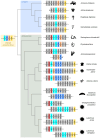
Figure 3.. Microsynteny of IL-1 genes in selected invertebrate phyla.
A schematic representation of the chromosomal regions surrounding the most conserved IL-1anc family paralog (IL-1, cyan) in selected invertebrates belonging to the Cnidaria and Ambulacraria and their phylogenetic relationships. Note that additional IL-1 genes exist outside these gene loci in several species, and that these genes were lost in corals. The IL-1s that were further characterized in subsequent experiments are numbered as they are referred to in the text instead of the generic IL-1 label. Selected genes encoding one-to-one orthologs present in multiple species are highlighted in color to emphasize the synteny between the genomic regions. An ancestral gene locus encoding both the IL-1 cytokine and its receptor (IL-1R) was inferred and is shown at the base of the phylogenetic tree.
Figure 4.
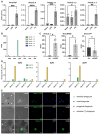
Figure 4.. Expression of IL-1 in invertebrates and jawless vertebrates.
(A) Three groups (N=4) of P. marinus ammocoetes were injected intraperitoneally with LPS or flagellin (Fla) or left untreated (naïve), and after 2 h the transcript levels of the indicated cytokines in the intestine/typhlosole were assessed by qRT-PCR. All levels were normalized to that of β-actin in the same sample and the mean ± SEM for each group is shown. A significant difference between the naïve and flagellin injected samples is indicated by asterisks (* p<0.05; ** p<0.01; *** p<0.001; **** p<0.0001, unpaired T-test). (B) The expression levels for each SpurIL-1 gene in the axial gland (axg), coelomocytes (coe), gut (gut), radial nerve (ran), ovary (ova), and testis (tes) were calculated from public RNAseq datasets (see Material and Methos for details). (C) The expression levels of N. vectensis IL-1anc (LOC5506646; NvecIL-1), NFκB (LOC5504072; NvecNFkB), and cytochrome p450 (LOC5501040; NvecCYP) in pools of untreated (ctrl, N=2) or 2’3’-cGAMP-treated (cGAMP, N=5) polyps were calculated from publicly-available RNAseq datasets (Margolis et al., 2021). A significant difference between the untreated and treated samples is indicated by asterisks (* p<0.05, unpaired T-test). (D) Three adult S. purpuratus (Sp#2, Sp#3, and Sp#4) were infected with V. diazotrophicus, and the expression levels of each SpurIL-1 gene were assessed by qRT-PCR prior to infection (t=0 h) and six hours later (t=6 h). All values were normalized to the levels of 18S transcripts in each sample, and the data for each individual is shown separately. (E) Fluorescence in situ hybridization was conducted on fixed coelomocytes (t=6 h) using the indicated HCR RNA probes with SpurIL-1 transcripts labelled in green. The nuclei were stained with DAPI (blue), and images were collected using visual light phase contrast (ph) and fluorescence microscopy. Representative images are shown separated by channel and merged (ph+green and blue+green). The phagocytes were classified manually based on their morphology (colored arrows).
Figure 5.
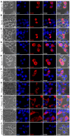
Figure 5.. Subcellular localization of IL-1s.
FLAG-tagged variants of the indicated full-length IL-1 proteins were transiently expressed in 293T cells, and their spatial distribution (shown in red) was visualized using immunofluorescence microscopy with a monoclonal anti-FLAG antibody. To classify the subcellular compartments, nuclei were stained with DAPI (blue), the cell morphology was visualized using phase contrast microscopy (ph), and images acquired in different channels were merged. Untransfected 293T cells served as negative controls.
Figure 6.
Figure 6.. Processing of IL-1s by caspase 1.
Human 293T cells were co-transfected with expression vectors for the indicated wildtype FLAG-tagged IL-1s (or mutants thereof in C), and V5-tagged Caspase 1 from humans (Hs, +), a catalytic mutant thereof (Δ), or Petromyzon marinus (Pm). Total cell lysates were resolved by SDS/PAGE and the proteins were visualized by Western blot using monoclonal anti-FLAG and anti-V5 antibodies, respectively. Non-transfected cells (NT) served as controls. The sizes of Caspase 1-dependent cleavage products are marked by a red asterisk on each blot. To illustrate that the lysates contained comparable concentrations of proteins, an anti-actin body was used. Theoretical sizes of the FLAG-tagged IL-1s are: PmarIL-1.1=33.0 kD (including signal peptide) or 26.9 kD (after cleavage of signal peptide); PmarIL-1.2=57.8 kD; PmarIL-1.3=47.9 kD; SpurIL-1.1=63.5 kD; SpurIL-1.2=55.2 kD; SpurIL-1.3=51.4 kD; mature SpurIL-1.3=30.6 kD; SpurIL-1.4=59.7 kD; SpIL-1.5=55.1 kD; HsapIL-1β=34 kD, mature HsapIL-1β-FLAG=20.6 kD.

References

    1. Beck G., O’Brien R. F., Habicht G. S., Stillman D. L., Cooper E. L., and Raftos D. A. (1993). Invertebrate cytokines. III: Invertebrate interleukin-1-like molecules stimulate phagocytosis by tunicate and echinoderm cells. Cell Immunol 146, 284–299. doi: S0008-8749(83)71027-0 [pii] 10.1006/cimm.1993.1027 - DOI - PubMed
    1. Bittrich S., Segura J., Duarte J. M., Burley S. K., and Rose Y. (2024). RCSB protein Data Bank: exploring protein 3D similarities via comprehensive structural alignments. Bioinformatics 40, btae370. doi: 10.1093/bioinformatics/btae370 - DOI - PMC - PubMed
    1. Boraschi D. (2022). What Is IL-1 for? The Functions of Interleukin-1 Across Evolution. Front Immunol 13, 872155. doi: 10.3389/fimmu.2022.872155 - DOI - PMC - PubMed
    1. Boraschi D., Italiani P., Weil S., and Martin M. U. (2018). The family of the interleukin-1 receptors. Immunol Rev 281, 197–232. doi: 10.1111/imr.12606 - DOI - PubMed
    1. Boulay J.-L., Du Pasquier L., and Cooper M. D. (2022). Cytokine Receptor Diversity in the Lamprey Predicts the Minimal Essential Cytokine Networks of Vertebrates. The Journal of Immunology 209, 1013–1020. doi: 10.4049/jimmunol.2200274 - DOI - PubMed

Publication types

LinkOut - more resources