Review
. 2025 Jul 28:34:102148.
doi: 10.1016/j.mtbio.2025.102148. eCollection 2025 Oct.

Organoid-on-a-chip (OrgOC): Advancing cystic fibrosis research

Affiliations
Review

Organoid-on-a-chip (OrgOC): Advancing cystic fibrosis research

Minjie Zheng et al. Mater Today Bio. .

Abstract

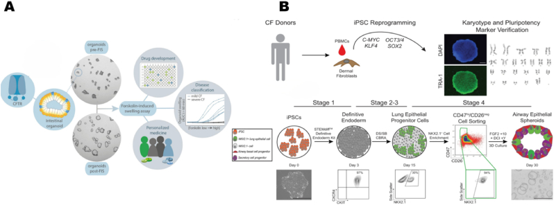
Cystic fibrosis (CF) is an autosomal recessive disorder resulting from impaired anion transport in the epithelium of multiple organs, thereby affecting various physiological functions throughout the body. The heterogeneity of CF complicates drug development, highlighting the growing importance of individualized therapies. CF patient-derived organoid models and organ-on-a-chip (OOC) platforms are promising in vitro models for recapitulating CF pathology, owing to their high simulation fidelity, individualized therapeutic capabilities, cost-effectiveness, and high-throughput screening potential. This review systematically summarizes the technological development pathways of patient-derived organoids and OOC platforms for CF, along with recent advances in their applications to CF-related basic research, and particularly focuses on exploratory studies using organoid-on-a-chip (OrgOC) systems to elucidate CF pathogenesis and assess therapeutic approaches.

Keywords: Cystic fibrosis; Organ-on-a-chip; Organoid; Organoid-on-a-chip.


Conflict of interest statement

The authors declare that they have no known competing financial interests or personal relationships that could have appeared to influence the work reported in this paper.

Figures

Image 1
Graphical abstract
Fig. 1
Fig. 1
A comparison is made between patients with cystic fibrosis and healthy individuals in the airways, digestive system, and sweat glands. Ion transport dysfunction, caused by mutations in the CFTR gene, leads to abnormal mucus accumulation in the Vater juxtaglomerular region of the airway and digestive systems, resulting in progressive mucus obstruction and secondary infections. This dysfunction also impairs the delivery of digestive enzymes, such as bile and pancreatic enzymes, to the small intestine, ultimately causing progressive nutrient malabsorption and metabolic disorders. In the sweat glands, functional CFTR proteins regulate electrolyte balance by facilitating chloride ion transport across membranes. However, defective CFTR proteins cause chloride ions to accumulate in sweat, where they combine with sodium ions to form a high concentration of salts, leading to the characteristic hypertonic, salty-smelling sweat seen in affected individuals.
Fig. 2
Fig. 2
Construction and application of cystic fibrosis organoids (A): The forskolin-induced swelling (FIS) assay transforms molecular functional defects in CFTR into quantifiable morphological changes in organoids, such as swelling degree, serving as a key bridge that connects gene-protein-function-phenotype in CF research. It can be applied in functional assessment, drug screening, and individualized diagnosis and treatment. Copyright 2020, Elsevier. (B): Construction of airway organoids derived from CF patients. First, somatic cells were reprogrammed into induced pluripotent stem cells (iPSCs), and their pluripotency and G-band karyotype labeling (TRA-1 staining and DAPI nuclear labeling) were then tested. A schematic diagram of redirected differentiation to generate airway epithelial cell spheres was examined using flow cytometry to ensure differentiation efficiency. Copyright 2022, Springer Nature.
Fig. 3
Fig. 3
lung-on-a-chip (A) Diagram of the Airway Chip illustrating the pseudostratified bronchial epithelium cultured under an air-liquid interface (ALI) on the top surface of a porous membrane, which separates the upper epithelial channel from the lower vascular channel containing primary human lung microvascular endothelial cells adhered to the bottom of the same membrane and exposed to dynamic fluid flow. Reproduced with permission [122]. Copyright 2022, Elsevier. (B) Design of the microfluidic chip, featuring a top block with space for Transwell housing and a bottom block composed of a fluidic channel and a central hexagonal pool. A gold electrode was integrated into the device, and the chip was mounted on a glass slide for stabilization. Images of the top and bottom blocks of the device, as well as the bonded chip, are shown. Reproduced with permission [123]Copyright 2023, ACS Publications.
Fig. 4
Fig. 4
Pancreas-on-a-chip, Microvascularized lung-on-a-chip (A) The single-channel chip, which mimics the structure of pancreatic ducts with branches and progressively narrower diameters, and the inset shows pancreatic ductal epithelial cells (PDECs) cultured on the chip; and the dual-channel pancreatic chip, which consists of two cell culture chambers and a porous membrane in which PDECs and islet cells are cultured on each side of the porous membrane. Reproduced with permission [125]. Copyright 2019 Springer Nature. (B) Structural diagram of a 96-well format microvascularized human lung-on-a-chip: top coverslips, vascular layer, membrane, airway layer, and bottomless 96-well. The vascular layer consists of human umbilical vein endothelial cells (HUVECs) and human lung fibroblasts (HLF), as well as normal human lung fibroblasts (NHLF), cultured in separate channels. The airway layer consists of normal human small airway epithelial cells (SAECs) cultured in the channel under S-ALI conditions. Reproduced with permission [126] Copyright 2020, The Royal Society of Chemistry.
Fig. 5
Fig. 5
Controlling the microenvironment of organoids-on-a-chip (A) Schematic diagram illustrating the experimental setup for generating tubular flow in a peristaltic gastric organoid chip. The actual figure of the gastric organoid chip consists of an incubation chamber, two reservoirs, and connections between two micropipettes. Reproduced with permission [130] Copyright 2018, The Royal Society of Chemistry. (B) The islet organoid chip comprises four layers: the top layer, the through-hole PDMS layer, the porous membrane, and the bottom layer; the differentiation and generation process of islet organoids derived from human induced pluripotent stem cells (hiPSCs) on the chip under three-dimensional perfusion culture conditions. The islet organoid differentiation process on the chip under three-dimensional perfusion culture conditions includes embryoid body (EB) formation, endoderm induction, and islet organoid differentiation. Reproduced with permission [131] Copyright 2021 Wiley.
Fig. 6
Fig. 6
Construction of microvascular organoids-on-a-chip (A) Assembly process of microvascularized organoid-on-a-chip: Human pluripotent stem cells (hPSCs) were differentiated into vascular cells and early neural organoids in suspension, and subsequently inoculated into the 3D-printed microfluidic chip. Physical diagram of the microvascularized organoid-on-a-chip platform. Reproduced with permission [133] Copyright 2022, The Royal Society of Chemistry. (B) Diagram of a high-throughput microvascularized organoid-on-a-chip system with ten microchannels, each controlled simultaneously by syringe pumps. Intra-channel structure featuring hydrogel containing endothelial cells attached to the channel walls, organoids captured in traps, and culture medium perfused through the channels via syringe pump for continuous flow. Reproduced with permission [136]. Copyright 2024 Springer Nature.

References

    1. Guo J., Garratt A., Hill A. Worldwide rates of diagnosis and effective treatment for cystic fibrosis. J. Cyst. Fibros. 2022;21:456–462. doi: 10.1016/j.jcf.2022.01.009. - DOI - PubMed
    1. Rommens J.M., Iannuzzi M.C., Kerem B.-S., Drumm M.L., Melmer G., Dean M., Rozmahel R., Cole J.L., Kennedy D., Hidaka N., Zsiga M., Buchwald M., Tsui L.-C., Riordan J.R., Collins F.S. Identification of the cystic fibrosis gene: chromosome walking and jumping. Science. 1989;245:1059–1065. doi: 10.1126/science.2772657. - DOI - PubMed
    1. Riordan J.R., Rommens J.M., Kerem B.-S., Alon N., Rozmahel R., Grzelczak Z., Zielenski J., Lok S., Plavsic N., Chou J.-L., Drumm M.L., Iannuzzi M.C., Collins F.S., Tsui L.-C. Identification of the cystic fibrosis gene: cloning and characterization of complementary DNA. Science. 1989;245:1066–1073. doi: 10.1126/science.2475911. - DOI - PubMed
    1. Levring J., Terry D.S., Kilic Z., Fitzgerald G., Blanchard S.C., Chen J. CFTR function, pathology and pharmacology at single-molecule resolution. Nature. 2023;616:606–614. doi: 10.1038/s41586-023-05854-7. - DOI - PMC - PubMed
    1. Jia S., Taylor-Cousar J.L. Cystic fibrosis modulator therapies. Annu. Rev. Med. 2023;74:413–426. doi: 10.1146/annurev-med-042921-021447. - DOI - PubMed

LinkOut - more resources