

Agreement between HRCT Imaging and Intraoperative Measurements in Predicting Stapedotomy Prosthesis Length in Otosclerosis Patients
- PMID: 40791838
- PMCID: PMC12335668
- DOI: 10.22038/ijorl.2025.81759.3749
Agreement between HRCT Imaging and Intraoperative Measurements in Predicting Stapedotomy Prosthesis Length in Otosclerosis Patients
Abstract
Introduction: This study aimed to evaluate the accuracy of preoperative high-resolution computed tomography (HRCT) imaging in measuring the distance from the long process of the incus to the footplate and its potential for predicting the optimal prosthesis length required for stapedotomy in patients with otosclerosis.
Materials and methods: This cross-sectional study included fifty patients scheduled for primary stapedotomy. A radiologist obtained and reconstructed preoperative HRCT scans of the temporal bone to measure the distance from the long process of the incus to the oval window in both axial and coronal views. These HRCT-derived measurements were then compared with intraoperative measurements performed by an otolaryngologist. The agreement between the two methods was assessed using correlation and Bland-Altman analysis.
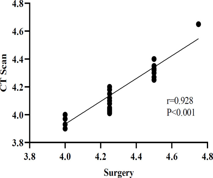
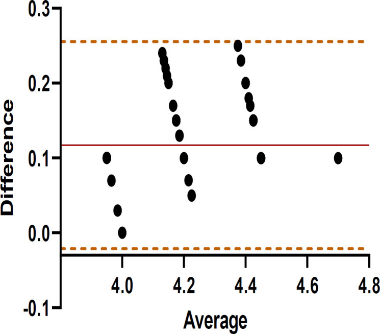
Results: The mean distances measured by HRCT and intraoperatively were 4.15mm and 4.27mm, respectively. A strong and statistically significant correlation (r=0.928, P<0.001) was observed between the two approaches, indicating a robust association. The Bland-Altman analysis revealed a mean bias of 0.11±0.07mm, with limits of agreement (LoAs) ranging from -0.02 to 0.26 mm, and no points exceeding the 95% LoAs. The maximum potential error between the two measurement methods was 0.28mm, suggesting that HRCT imaging can reliably predict prosthesis length. In a stratified analysis based on the surgical distance (≤4 mm [N=11], 4.25mm [N=25], ≥4.5mm [N=13]), good agreement was maintained in the Bland-Altman analysis.
Conclusion: Preoperative HRCT imaging may be a valuable tool for accurately predicting the required prosthesis length prior to stapedotomy in otosclerosis patients.
Keywords: CT scan; Otosclerosis; incus bone; stapedotomy.
©2025 Mashhad University of Medical Sciences.
Conflict of interest statement
The authors declare no conflicts of interest regarding the publication of this paper.
Figures


References
-
- Declau F, Van Spaendonck M, Timmermans J, Michaels L, Liang J, Qiu J, et al. Otosclerosis and Stapes Surgery. Karger Publishers; 2007. Prevalence of histologic otosclerosis: an unbiased temporal bone study in Caucasians; pp. 6– 16. - PubMed
-
- Parving YS. Clinical otosclerosis, prevalence estimates and spontaneous progress. Acta oto-laryngologica. 1999;119(4):468 –72. - PubMed
-
- Eza-Nuñez P, Manrique-Rodriguez M, Perez-Fernandez N. Otosclerosis among patients with dizziness. Rev Laryngol Otol Rhinol (Bord) 2010;131(3):199–206. - PubMed
-
- Uppal S, Bajaj Y, Rustom I, Coatesworth AP. Otosclerosis 1: the aetiopathogenesis of otosclerosis. Int J Clin Pract. 2009;63(10):1526–30. - PubMed
LinkOut - more resources
Full Text Sources