. 2025 Jun 19;2(3):100130.
doi: 10.1016/j.bneo.2025.100130. eCollection 2025 Aug.

GCK inhibition enhances iberdomide antimyeloma effects by promoting IKZF1 degradation via a CRBN-independent mechanism

Affiliations

GCK inhibition enhances iberdomide antimyeloma effects by promoting IKZF1 degradation via a CRBN-independent mechanism

Shirong Li et al. Blood Neoplasia. .

Abstract

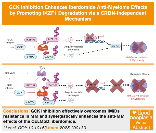
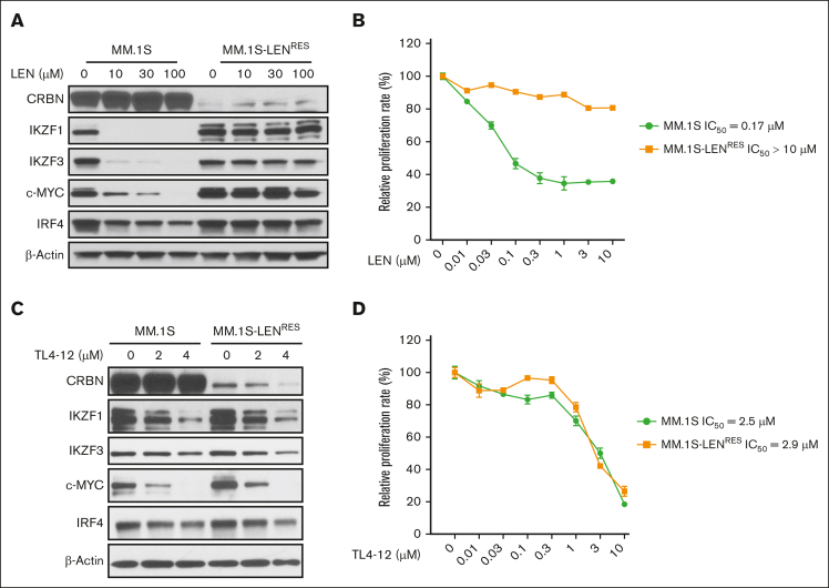
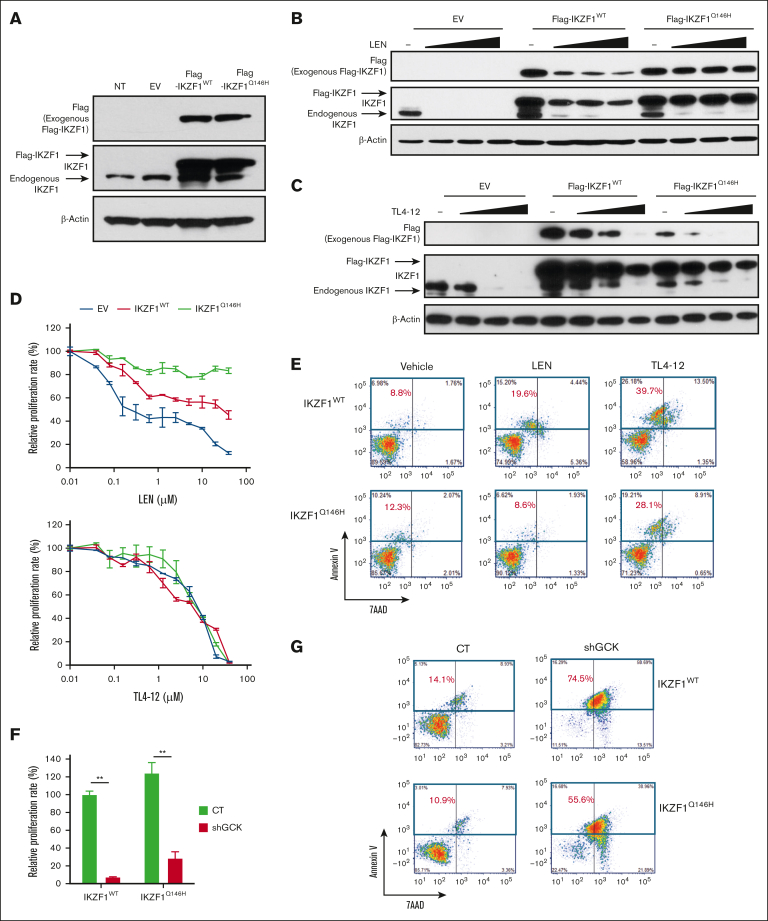
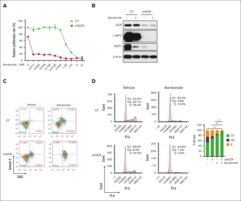
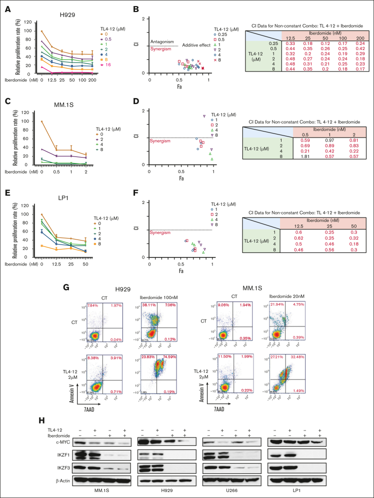
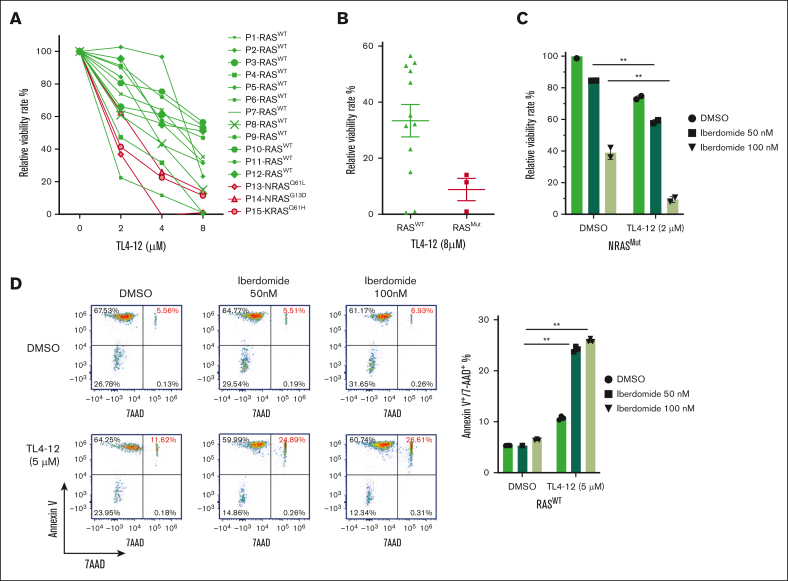
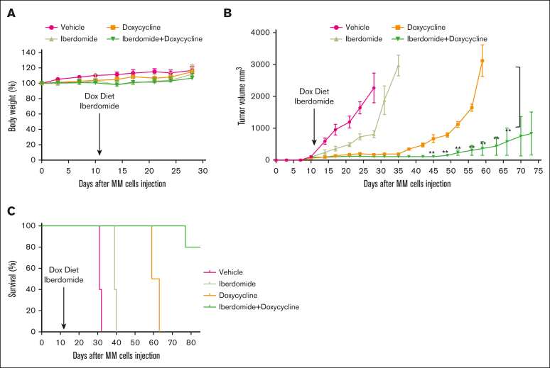
Our recent study identifies germinal center kinase (GCK) as a novel therapeutic target in RAS-mutated multiple myeloma (MM). Inhibiting GCK downregulates critical transcriptional factors, notably IKZF1/3, BCL-6, and c-MYC proteins, leading to MM cell growth inhibition and cell death. Distinct from immunomodulatory drug (IMiD)-induced IKZF1/3 degradation, GCK inhibition triggers IKZF1/3 proteolysis through a cereblon (CRBN) E3 ligase-independent mechanism. Here, we demonstrated that GCK inhibition overcomes IMiD resistance in MM. An isogenic subline of MM.1S cells with acquired lenalidomide resistance remains sensitive to GCK inhibition-induced IKZF1/3 downregulation and cell growth inhibition. Consistently, the CRBN-resistant IKZF1 Q146H mutant maintains sensitivity to GCK inhibitor-induced degradation, similar to the IKZF1 wild-type protein, suggesting a CRBN-independent protein degradation. In accordance with the distinct IKZF1/3 degradation mechanisms, GCK silencing enhances iberdomide-induced IKZF1/3 and c-MYC downregulation and MM growth inhibition. More importantly, the combination of a GCK inhibitor with iberdomide exhibited synergistic anti-MM effects in a panel of MM cell lines and primary plasma cells. The synergistic effects were confirmed in an MM xenograft mouse model, in which combining GCK silencing and iberdomide resulted in significantly enhanced tumor inhibition and prolonged mice survival compared to single treatments. These findings underscore GCK as a promising therapeutic target for bypassing IMiD resistance in MM. Combining GCK inhibition with iberdomide could provide a novel strategy to manage relapsed or refractory patients with multidrug resistance, especially after the exhaustion of immunotherapy.


Conflict of interest statement

Conflict-of-interest disclosure: S. Li is currently a full-time employee of Bristol Myers Squibb. C.M. is a full-time employee of Sanofi. S. Lentzsch reports Caelum Biosciences patents and royalties on CAEL-101; advisory board fees from Pfizer, GlaxoSmithKline, Karyopharm, Regeneron, Janssen, and Sanofi; honoraria from Regeneron, PeerView, Clinical Care Options, Medscape, and Springer Healthcare; membership on an entity's board of directors or advisory committees for Regeneron, Janssen, and Bristol Myers Squibb; and research funding from Sanofi and Zentalis. The remaining authors declare no competing financial interests.

Figures

None
Graphical abstract
Figure 1.
Figure 1.
GCK inhibition overcomes LEN resistance in MM cells. (A) MM.1S and MM.1S-LENRES were treated with LEN at the indicated concentrations for 24 hours. CRBN, IKZF1, c-MYC, IKZF3, and IRF4 protein levels in cell lysates were examined by western blotting. (B) MM.1S and MM.1S-LENRES were treated with LEN at the indicated concentrations for 3 days for cell proliferation detected by AQueous One Solution Cell Proliferation Assay (MTS). (C) MM.1S and MM.1S-LENRES were cultured in the presence of TL4-12 at the indicated concentrations for 24 hours to analyze CRBN, IKZF1, c-MYC, IKZF3, and IRF4 expression by western blotting. (D) MM.1S and MM.1S-LENRES were treated with TL4-12 at the indicated concentrations for 3 days for cell proliferation detected as in panel B. IC50, 50% inhibitory concentration.
Figure 2.
Figure 2.
GCKi induces anti-MM effects by targeting IKZF1 via a CRBN-independent mechanism. MM.1S-tet-on-shGCK cells were infected by PCDH-Flag-IKZF1-WT (IKZF1WT) or PCDH-Flag-IKZF1-Q146H (IKZF1Q146H) lentivirus and sorted by green fluorescent protein after 3 days. (A) Cell lysates were analyzed by western blotting to confirm IKZF1WT and IKZF1Q146H expression. (B-C) Sorted cells were treated with LEN at 2, 4, or 8 μM (B) or TL4-12 at 5, 10, 15 μM (C) or dimethyl sulfoxide (DMSO; 0.01%) for 24 hours. Cell lysates were analyzed by western blotting. (D) Sorted cells were treated with LEN or TL4-12 at indicated concentrations for 5 days. Cell proliferation was detected by MTS. (E) Sorted cells were treated with 5-μM LEN, 5-μM TL4-12, or DMSO (0.01%) for 5 days. Treated cells were stained with Annexin V and 7-AAD for apoptosis analysis. (F-G) Sorted cells were treated with 400 ng/mL doxycycline (DOX) for 5 days to induce GCK knockdown (shGCK) or DMSO (0.01%) as control (CT). Cell proliferation was detected by MTS (F). ∗∗P < .01 (by 2-way analysis of variance [ANOVA]). Apoptosis assay was detected by Annexin V and 7-AAD staining (G).
Figure 3.
Figure 3.
The combination of GCK silencing and iberdomide shows enhanced anti-MM effects. (A) DOX-inducible shGCK MM.1S cells were treated with 400 ng/mL DOX for 48 hours, then treated with different doses of Iberdomide for 72 hours. Cell proliferation was detected using MTS. (B) DOX-inducible shGCK MM.1S cells were treated with 400 ng/mL DOX for 48 hours to induce GCK knockdown (shGCK), then treated with 1 nM iberdomide for 24 hours. The protein expression level of GCK, c-MYC, and IKZF1 were detected by western blot using β-actin as a loading CT. (C-D) DOX-inducible shGCK MM.1S cells were treated with 400 ng/mL DOX for 2 days to induce GCK knockdown (shGCK), then treated with 1 nM iberdomide for 5 days. Cell apoptosis was detected by flow cytometry after Annexin V and 7-AAD staining (C); cell cycle was analyzed by flow cytometry after PI staining (D). ∗∗P < .01 (by 1-way ANOVA).
Figure 4.
Figure 4.
The combination of GCKi and iberdomide synergistically exerts anti-MM effects. (A) H929 cells proliferation was assessed after treatment with the combination of TL4-12 and iberdomide at indicated doses for 4 days. (B) The CI value was analyzed by CompuSyn software. (C-D) MM.1S cells proliferation was assessed and the combination of TL4-12 and iberdomide at indicated doses for 4 days (C), and CI value was analyzed by CompuSyn (D). (E-F) LP1 cells proliferation was assessed after treatment with the combination of TL4-12 and iberdomide at indicated doses for 4 days (E), and CI value was analyzed by CompuSyn (F). (G) H929 and MM.1S cells were treated with TL4-12 and iberdomide at the indicated concentrations for 4 days. Cell apoptosis was detected by flow cytometry after Annexin V and 7-AAD staining. (H) MM cells were treated with TL4-12 (MM.1S and U266, 5 μM; H929 and LP-1, 2 μM) and/or iberdomide (MM.1S and U266, 1 nM; H929 and LP-1, 30 nM) for 24 hours. The protein expression level of c-MYC, IKZF3, and IKZF1 were detected by western blot using β-actin as a loading CT. GCKi is GCK inhibition.
Figure 5.
Figure 5.
GCKi and iberdomide show synergistic anti-MM effects on primary MM. (A) Primary MM samples (RASWT, n = 12; RASMut, n = 3) were cocultured with bone marrow stromal cells and treated with TL4-12 at the indicated concentrations for 5 days. Cell viability rate was analyzed by MTS. (B) Primary MM cell viability inhibition rate by TL4-12 at 8 μM were compared between RASWT and RASMut. (C) NRASG61K primary CD138+ MM cells cocultured with bone marrow stromal cells were treated with TL4-12 and iberdomide at the indicated concentrations for 4 days. Cell viability was analyzed by MTS. (D) RASWT plasma CD138+ cells were treated with TL4-12 and iberdomide at the indicated concentrations for 4 days. Cell apoptosis was detected by flow cytometry after Annexin V and 7-AAD staining.
Figure 6.
Figure 6.
GCK silencing and iberdomide combination enhances anti-MM effects on MM xenograft model. Tet-on-sh-GCK-MM1.S cells were subcutaneously injected into severe combined immunodeficient beige mice. (A) Body weights were monitored every 3 days. No significant differences were observed between the different groups. (B) Subcutaneous tumor growth was measured using calipers and calculated with the volume formula: 0.5 × long diameter × (short diameter)2 for up to 75 days. Each bar represents the mean ± standard error of the mean (n = 5). ∗∗P < .01. (C) Kaplan-Meier survival analysis for the MM tumor model during 6 weeks of follow-up (n = 5 per group). Using a Log-Rank test, a survival benefit was observed for DOX vs vehicle (∗P < .05) and iberdomide + DOX vs vehicle (∗P < .05).

Similar articles

References

    1. Kumar SK, Rajkumar V, Kyle RA, et al. Multiple myeloma. Nat Rev Dis Primers. 2017;3 - PubMed
    1. Hamadeh IS, Friend R, Mailankody S, Atrash S. Chimeric antigen receptor T-cells: a review on current status and future directions for relapsed/refractory multiple myeloma. Front Oncol. 2024;14 - PMC - PubMed
    1. Ledergor G, Fan Z, Wu K, et al. CD4+ CAR T-cell exhaustion associated with early relapse of multiple myeloma after BCMA CAR T-cell therapy. Blood Adv. 2024;8(13):3562–3575. - PMC - PubMed
    1. Holstein SA, Grant SJ, Wildes TM. Chimeric antigen receptor T-cell and bispecific antibody therapy in multiple myeloma: moving into the future. J Clin Oncol. 2023;41(27):4416–4429. - PMC - PubMed
    1. Lee H, Ahn S, Maity R, et al. Mechanisms of antigen escape from BCMA- or GPRC5D-targeted immunotherapies in multiple myeloma. Nat Med. 2023;29(9):2295–2306. - PMC - PubMed

LinkOut - more resources