. 2025 Jul 28:12:1616186.
doi: 10.3389/fvets.2025.1616186. eCollection 2025.

Astragalus polysaccharides protect against Di-n-butyl phthalate-induced testicular damage by modulating oxidative stress, apoptosis, and the PI3K/Akt/mTOR pathway in rats

Affiliations

Astragalus polysaccharides protect against Di-n-butyl phthalate-induced testicular damage by modulating oxidative stress, apoptosis, and the PI3K/Akt/mTOR pathway in rats

Manal R Bakeer et al. Front Vet Sci. .

Abstract

Introduction: Di-n-butyl phthalate (DBP), a common plasticizer, is associated with oxidative stress and male reproductive toxicity. Astragalus polysaccharides (APS) have known antioxidative and anti-inflammatory properties, but their role in male reproductive health has not been fully elucidated.

Methods: Twenty-four male rats were randomly assigned to four groups (n = 6 each): control, DBP-only (500 mg/kg/day), APS-only (200 mg/kg/day), and APS + DBP (500 mg/kg/day DBP + 200 mg/kg/day APS). Treatments were administered orally for 8 weeks. Biochemical, histological, and molecular analyses were conducted to evaluate testicular function, oxidative stress markers, and gene expression.

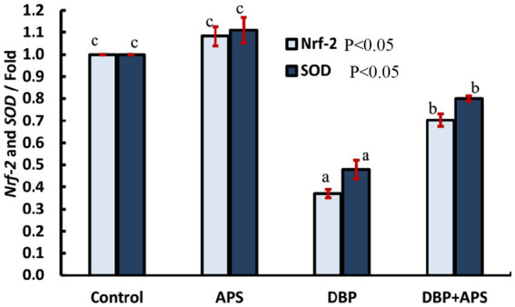
Results: DBP exposure significantly decreased serum testosterone levels, catalase (CAT) activity, lactate dehydrogenase (LDH) activity, and sperm quality, while increasing malondialdehyde (MDA) levels and apoptotic markers Casp3, Casp9. APS co-treatment significantly restored antioxidant enzyme activity, improved sperm parameters, reduced MDA levels, and alleviated histopathological damage. Gene expression analysis revealed upregulation of Nrf2 and SOD, and modulation of the PI3K/AKT/mTOR signaling pathway.

Discussion: APS exerts protective effects against DBP-induced testicular damage by enhancing antioxidant defenses and regulating key molecular pathways. These findings highlight the therapeutic potential of APS in preventing male infertility associated with environmental toxicants.

Keywords: PI3K/AKT/mTOR pathway; apoptosis; astragalus polysaccharides; dibutyl phthalate; male infertility; oxidative stress; reproductive toxicity.


Conflict of interest statement

The authors declare that the research was conducted in the absence of any commercial or financial relationships that could be construed as a potential conflict of interest.

Figures

Figure 1
Figure 1
GC–MS chromatogram showing various peaks and their retention times for Astragalus membranaceus.
Figure 2
Figure 2
The percentage of free radical scavenging activity of APS with DPPH against that of ascorbic acid.
Figure 3
Figure 3
Mean serum testosterone concentrations (ng/ml) in adult male albino rats treated with DBP and APS with error bars. Different superscript letters (a,b,c) indicate significance at p < 0.05.
Figure 4
Figure 4
Mean percentage of sperm motility, viability, and abnormality in adult male albino rats treated with DBP and APS with error bars. Different superscript letters (a, b, c, d) indicate significance at p < 0.05.
Figure 5
Figure 5
Mean sperm concentration (x106/ml) in adult male albino rats treated with DBP and APS with error bars. Different superscript letters (a, b, c) indicate significance at p < 0.05.
Figure 6
Figure 6
Mean testicular lactate dehydrogenase activity (LDH IU/g) in adult male albino rats treated with DBP and APS with error bars. Different superscript letters (a,b,c) indicate significance at p < 0.05.
Figure 7
Figure 7
Mean testicular malondialdehyde levels (MDA nmol/g) in adult male albino rats treated with DBP and APS with error bars. Different superscript letters (a, b, c) indicate significance at p < 0.05.
Figure 8
Figure 8
Mean testicular Catalase activity (U/g) in adult male albino rats treated with DBP and APS with error bars. Different superscript letters (a, b, c) indicate significance at p < 0.05.
Figure 9
Figure 9
mRNA expression of the INSL3 gene in testicular tissue of adult male albino rats treated with DBP and APS with error bars. Different superscript letters (a, b, c) indicate significance at p < 0.05.
Figure 10
Figure 10
mRNA expression of antioxidant-related genes: Nrf-2 and SOD in testicular tissue of adult male albino rats treated with DBP and APS with error bars. Different superscript letters (a, b, c) indicate significance at p < 0.05.
Figure 11
Figure 11
mRNA expression of the NBN gene in testicular tissue of adult male albino rats treated with DBP and APS with error bars. Different superscript letters (a,b,c) indicate significance at p < 0.05.
Figure 12
Figure 12
mRNA expression of apoptosis-related genes: Casp3 and Casp9 genes in testicular tissue of adult male albino rats treated with DBP and APS with error bars. Different superscript letters (a, b, c) indicate significance at p < 0.05.
Figure 13
Figure 13
mRNA expression of PI3K/AKT signaling-related Casp9 genes in testicular tissue of adult male albino rats treated with DBP and APS with error bars. Different superscript letters (a, b, c) indicate significance at p < 0.05.
Figure 14
Figure 14
(a–f) Hematoxylin and eosin (H&E) stained testicular sections from adult male albino rats. (a) Control and (b) APS group showing normal active seminiferous tubule with spermatogenic cells; spermatogonia (black arrow), primary spermatocyte (yellow arrow), secondary spermatocyte (red arrow), spermatid (blue arrow), Sertoli cells (blue circle) with lumen filled with sperms (yellow star). Normal interstitial tissue containing Leydig cells (blue circle; X400). Testicular sections from DBP group (c) showed degeneration, necrosis (blue arrow), and pyknosis of spermatogenic cells with sloughing into the lumen (red arrow), which appeared devoid of sperm (black star). Germinal epithelial cells could not be differentiated within the wall (black arrow). (d) Degenerated seminiferous tubules appeared irregular in shape (black arrow) with widened interstitial spaces (edema; red arrow) and necrosis of Leydig cells (blue chevron; X400). (e) Spermatogenic cells displayed ballooning and vacuolization (red arrow), degeneration (black arrow) with necrotic debris in the tubular lumina (blue chevron), and absence of sperms (black star; X100). (f) DBP + APS group exhibited some nearly normal seminiferous tubules with several layers of spermatogenic cells, spermatogonia (black arrow), primary spermatocytes (yellow arrow), with Sertoli cell (blue circle), and the lumen contained sperms (yellow star). Narrow interstitial tissue, no edematous area (black star), and nearly normal Leydig cells (blue arrow; X400).
Figure 15
Figure 15
(a–f) Immunohistochemically caspase-3-stained testicular sections from adult male albino rats. (a) Control and (b) APS group exhibiting negative immune reactivity to caspase-3 (x400), DBP-exposed group (c–e) shows strong positive immunoreaction(x400). (e) The interstitial tissue edema (yellow arrow; x100). (f) DBP+APS group shows a negligible immune reaction with several layers of spermatogenic cells (x400).

References

    1. Pan J, Liu P, Yu X, Zhang Z, Liu J. The adverse role of endocrine disrupting chemicals in the reproductive system. Front Endocrinol. (2024) 14:1324993. doi: 10.3389/fendo.2023.1324993, PMID: - DOI - PMC - PubMed
    1. Yan Y, Bender ML, Brook EJ. Two-million-year-old snapshots of atmospheric gases from Antarctic ice. Nature. (2019) 574:663–6. doi: 10.1038/s41586-019-1692-3, PMID: - DOI - PubMed
    1. Buzadzic B, Vucetic M, Jankovic A, Stancic A, Korac A, Korac B, et al. New insight into male fertility: the importance of NO. Br J Pharmacol. (2015) 172:1455–67. doi: 10.1111/bph.12675 - DOI - PMC - PubMed
    1. Soliman SS, El-Sheikh M, Taha D A, et al. Expression of growth factors in buffalo ovarian tissue across different follicular developmental stages. Arch Gynecol Obstet. (2025) 303: 1–11. doi: 10.1007/s00404-025-08090-8 - DOI - PubMed
    1. Soliman S, El-Sanea M, Kandil M, Aboelmaaty M, Abdoon S. Impact of reproductive status, body condition score, and locality on hormonal, and some blood metabolites in Egyptian buffaloes. Egypt J Vet Sci. (2024) 55:1387–96. doi: 10.21608/ejvs.2024.252235.1699 - DOI

LinkOut - more resources