. 2025 Jul 28:15:1621846.
doi: 10.3389/fcimb.2025.1621846. eCollection 2025.

Drosophila melanogaster as a model system for studying the effects of porcine rotavirus on intestinal immunity

Affiliations

Drosophila melanogaster as a model system for studying the effects of porcine rotavirus on intestinal immunity

Xiao Chun Wang et al. Front Cell Infect Microbiol. .

Abstract

Introduction: Drosophila melanogaster is a quintessential model organism that has been used in many scientific studies. The intestinal immune response of flies is a critical component of their innate immune system. Given that flies primarily consume decaying organic matter, harmful microorganisms present in their food can enter the intestine, leading to frequent infections by exogenous pathogens. When these pathogens are introduced into the intestinal environment, a cascade of immune responses is triggered within the intestinal tissue, aimed at preserving the integrity of the intestinal barrier and ensuring the proper physiological functions of the gut. Porcine rotavirus (PoRV) is a key pathogen that causes diarrhea in pigs, and PoRV infection can significantly reduce piglet survival rates.

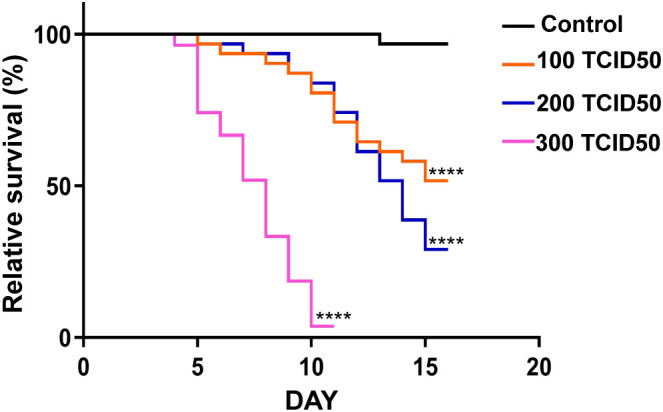
Methods: In this study, wild-type flies were orally administered PoRV to establish an effective intestinal damage animal model, and a detailed investigation of the antiviral immune defense mechanism in the fly intestine was performed.

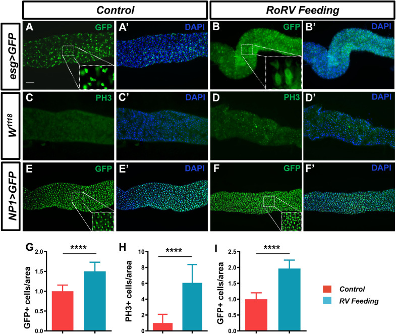
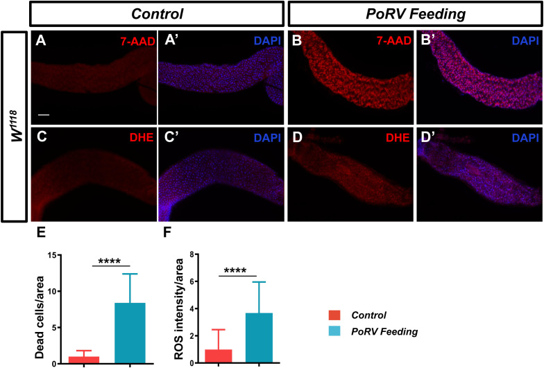
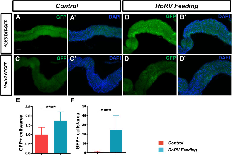
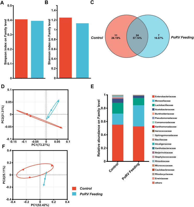
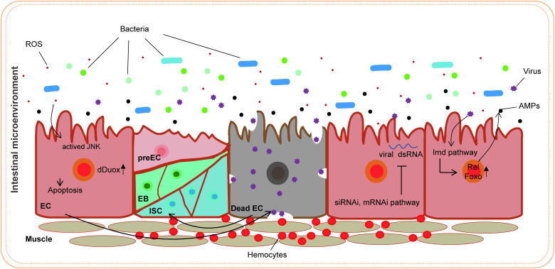
Results and discussion: Our study revealed that PoRV infection caused a reduction in the survival rate of flies and an increase in intestinal epithelial cell death. Concurrently, PoRV infection significantly promoted the proliferation and differentiation of intestinal cells, contributing to the maintenance of intestinal homeostasis. After the activation of JAK/STAT signaling in the intestines of infected Drosophila, there was an increase in the levels of reactive oxygen species (ROS). This elevation was concomitant with the release of antimicrobial peptides (AMPs), which play a crucial role in pathogen clearance. Additionally, we identified substantial aggregation of hemocytes in the midgut. The composition of the intestinal microbiota also underwent changes, potentially playing a role in intestinal immune defense. Moreover, PoRV can evade clearance via the RNA interference (RNAi) pathway. In summary, PoRV infection in the fly intestine activates multiple immune defense mechanisms to eliminate the pathogen, offering a theoretical basis for PoRV prevention and control.

Keywords: D. melanogaster; JAK/STAT signaling; intestinal immunity; pathogen; porcine rotavirus (PoRV).


Conflict of interest statement

The authors declare that the research was conducted in the absence of any commercial or financial relationships that could be construed as a potential conflict of interest.

Figures

Figure 1
Figure 1
Survival rates of the control and experimental groups of Drosophila. W1118 adult flies were cultured in standard medium or medium supplemented with PoRV. These experiments were conducted in vials, each containing 15 male and 15 female flies. Control, sucrose (5%, w/v); experimental groups, RoRV (5% sucrose plus the 100 TCID50, 200 TCID50 or 300 TCID50 values of RoRV). At least three replicates were performed for each treatment. Survival differences were analyzed via the log-rank test. ****P <0.0001 vs. the control group.
Figure 2
Figure 2
PoRV treatment changes the number of intestinal cells in the Drosophila gut. Three- to five-day-old esg>GFP, W1118 or NP1>GFP adult flies were fed sucrose or 300 TCID50 of RoRV plus 5% sucrose for 72 h. (A, A’, B, B’) Progenitor cells were stained with anti-GFP antibodies (green). (C, C’, D, D’) The proliferation of ISCs in the posterior midgut was evaluated with anti-PH3 antibodies (green). (E, E’, F, F’) EC cells were stained with anti-GFP antibodies (green). DAPI, nuclei (blue). (H) Quantification of the number of PH3+ cells per unit area of the midgut in C and D, n > 15. (G, I) The number of GFP+ cells per unit area of the midgut in A, B and E, F are shown, n>15. Scale bars: 50 μm.
Figure 3
Figure 3
PoRV induced cell death and increased ROS levels. Three- to five-day-old W1118 adult flies were fed 5% sucrose or 300 TCID50 of RoRV plus 5% sucrose for 72 h. (A, A’, B, B’) Dead cells were detected with 7-AAD. (C, C’, D, D’) ROS levels in the adult fly midgut were evaluated via DHE staining. (E) Quantification of the number of dead cells per unit area of the midgut in A and B, n > 12. (F) Quantification of DHE intensity per unit area of the midgut in C and D, n > 10. Scale bars: 50 μm.
Figure 4
Figure 4
PoRV feeding activated the JAK/STAT pathway and increased midgut hemocyte numbers. (A, A’, B, B’) JAK/STAT pathway activity was assessed using the 10XSTAT-GFP reporter. The number of GFP+ cells was greater in the flies infected with PoRV than in the uninfected flies. (C, C’, D, D’) High ROS levels mediated by PoRV led to the aggregation of hemocytes. A hml>2XEGFP transgene was used to label hemocytes. (E, F) The numbers of GFP+ cells per unit area of the midgut in A, B and C, D are shown, n > 15. Scale bars: 50 μm. ****P < 0.0001, scale bars: 50 μm.
Figure 5
Figure 5
qRT-PCR analysis of antimicrobial peptides (AMPs) and siRNA/miRNA pathway member levels in the adult gut or body. W1118 adult male flies that were treated with 300 TCID50 and their guts were analyzed. (A) Relative gene expression of AMPs in the gut. (B, C) Relative expression of genes associated with the siRNA/miRNA pathways in the gut and body. Similar expression patterns were observed in three independent experiments. AttA, Attacin A; Dpt, Diptericin; CecA1, Cecropin A1; Dro, Drsosocin A; Def, Defensin; Drs, Drosomycin; Mtk, Metchnikowin.
Figure 6
Figure 6
PoRV infection affects the gut microbiota composition. (A, B) α diversity. The bar plot analysis shows the biodiversity values for the Simpson and Shannon indices. No statistically relevant differences were observed. (C) Venn diagram showing the number of families unique to and shared among different groups. (D, F) Principal coordinate analysis (PCoA) for both UniFrac distances. (Student’s t test: weighted P = 0.386; unweighted P = 0.406). (E) Bacterial composition of the control and PoRV groups. Relative taxonomic abundances are shown at the family level. All bacterial taxa present at < 1% relative abundance were grouped into the “Other” classification.
Figure 7
Figure 7
Antiviral strategies in the D. melanogaster intestine.

Similar articles

References

    1. Amcheslavsky A., Jiang J., Ip Y. T. (2009). Tissue damage-induced intestinal stem cell division in Drosophila . Cell Stem Cell 4, 49–61. doi: 10.1016/j.stem.2008.10.016, PMID: , PMID: - DOI - PMC - PubMed
    1. Arch M., Vidal M., Koiffman R., Melkie S. T., Cardona P.-J. (2022). Drosophila melanogaster as a model to study innate immune memory. Front. Microbiol. 13. doi: 10.3389/fmicb.2022.991678, PMID: , PMID: - DOI - PMC - PubMed
    1. Buchon N., Silverman N., Cherry S. (2014). Immunity in Drosophila melanogaster–from microbial recognition to whole-organism physiology. Nat. Rev. Immunol. 14, 796–810. doi: 10.1038/nri3763, PMID: , PMID: - DOI - PMC - PubMed
    1. Chtarbanova S., Lamiable O., Lee K.-Z., Galiana D., Troxler L., Meignin C., et al. (2014). Drosophila C virus systemic infection leads to intestinal obstruction. J. Virol. 88, 14057–14069. doi: 10.1128/jvi.02320-14, PMID: , PMID: - DOI - PMC - PubMed
    1. Costa A., Jan E., Sarnow P., Schneider D. (2009). The Imd pathway is involved in antiviral immune responses in Drosophila . PloS One 4, e7436–e7436. doi: 10.1371/journal.pone.0007436, PMID: , PMID: - DOI - PMC - PubMed

Substances

LinkOut - more resources