. 2025 Jul 30;14(7):4357-4368.
doi: 10.21037/tcr-2024-2217. Epub 2025 Jul 22.

miR-490-5p inhibits the progression of osteosarcoma by targeting HDAC2

Affiliations

miR-490-5p inhibits the progression of osteosarcoma by targeting HDAC2

Huiqun Jiang et al. Transl Cancer Res. .

Abstract

Background: Osteosarcoma (OS) is the most prevalent malignant bone tumor and has a particularly unfavorable prognosis. Although miR-490-5p is regarded as an established diagnostic and predictive marker for human cancers, the role of miR-490-5p in OS remains presently unclear. The aim of this study was to clarify the function of miR-490-5p in OS.

Methods: Bioinformatics analysis of OS cells was performed to identify differentially expressed microRNAs (miRNAs, miRs). Then, the expression profiles of miR-490-5p in various OS cells were determined by real-time polymerase chain reaction analysis. The roles of miR-490-5p in OS cells were assessed through in vitro cytological experiments. Bioinformatics methods were used to predict target genes. The relationship between miR-490-5p and HDAC2 was demonstrated by a dual-luciferase reporter gene assay.

Results: Expression of miR-490-5p was relatively decreased in OS cells. Overexpression of miR-490-5p inhibited proliferation and metastasis of OS cells. Moreover, miR-490-5p was found to negatively regulate HDAC2 as a downstream target gene. Recovery experiments confirmed that HDAC2 overexpression rescued the inhibitory effect on OS progression by overexpression of miR-490-5p.

Conclusions: MiR-490-5p directly regulated HDAC2 expression, thereby modulating the growth and metastatic capacity of OS cells. The miR-490-5p/HDAC2 axis could serve as a promising therapeutic target for OS.

Keywords: HDAC2; Osteosarcoma (OS); metastasis; miR-490-5p; proliferation.


Conflict of interest statement

Conflicts of Interest: All authors have completed the ICMJE uniform disclosure form (available at https://tcr.amegroups.com/article/view/10.21037/tcr-2024-2217/coif). The authors have no conflicts of interest to declare.

Figures

Figure 1
Figure 1
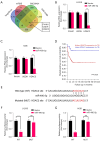
Expression of miR-490-5p was downregulated in OS tissues and cell lines. (A) Volcano plot of differentially expressed miRNAs (P<0.05 and |log2 fold change| ≥1). (B) Heat map of differentially expressed miRNAs in the GSE28423 dataset from the GEO database. (C) Expression profiles of miR-490-5p in normal and OS tissue specimens retrieved from the GSE28423 dataset. (D) Representative FISH images of the expression patterns of miR-490-5p in human OS tissues. Blue signal: DAPI staining; red signal: miR-490-5p probe. Scale bar =50 µm, inset =50 µm. (E) Statistical analysis of FISH results based on the expression level of miR-490-5p in T1 (n=9) and T2 (n=31) stages in OS tissues. (F) Expression levels of miR-490-5p of U-2OS, Saos-2, 143B, and MNNG-HOS relative to hFOB1.19 cells were determined by qRT-PCR analysis. The results are presented as the mean ± SD. **, P<0.01; ***, P<0.001. DAPI, 4',6-diamidino-2-phenylindole; FISH, fluorescence in situ hybridization; GEO, Gene Expression Omnibus; OS, osteosarcoma; qRT-PCR, quantitative reverse transcription polymerase chain reaction; SD, standard deviation.
Figure 2
Figure 2
miR-490-5p inhibits OS progression in vitro. (A) The efficiency of miR-490-5p overexpression in U-2OS and 143B cells was determined by qRT-PCR analysis. (B,C) The CCK-8 assay was used to determine the effect of miR-490-5p overexpression on the proliferative capacity of OS cells. (D,E) The colony formation assay was used to assess the effects of miR-490-5p overexpression on colony formation ability of OS cells (crystal violet staining). (F,G) Representative wound healing photomicrographs and quantitative analysis of OS cell mobility at 0 and 48 h. Scale bar =100 µm. (H,I) The invasive capacity of OS cells was determined based on the presence or absence of miR-490-5p overexpression (crystal violet staining). Scale bar =50 µm. The results are presented as the mean ± SD. ***, P<0.001. CCK-8, Cell Counting Kit-8; OS, osteosarcoma; qRT-PCR, quantitative reverse transcription polymerase chain reaction; SD, standard deviation.
Figure 3
Figure 3
HDAC2 is a direct target of miR-490-5p. (A) A Venn diagram showing bioinformatics analysis of miR-490-5p target genes. (B,C) The mutual putative target genes expression in OS cells transfected with a negative control or miR-490-5p plasmid. (D) Kaplan-Meier analysis of the overall survival rate of OS patients related to HDAC2 expression (n=88) was generated using the R2 Genomics Analysis and Visualization Platform. (E) HDAC2 3'-UTR contains one predicted miR-490-5p binding site. (F,G) miR-490-5p significantly inhibited luciferase activity of the 3'-UTR wild-type reporter of HDAC2, but not the mutant. The results are presented as the mean ± SD. ***, P<0.001; ns, P>0.05. MUT, mutant; OS, osteosarcoma; SD, standard deviation; WT, wild-type.
Figure 4
Figure 4
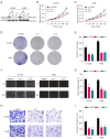
HDAC2 promotes progression and metastasis of OS cells. (A) The knockdown efficiency of HDAC2 in OS cells was confirmed by western blot analysis. (B,C) The CCK-8 assay demonstrated that HDAC2 significantly suppressed proliferation of OS cells. (D,E) The effect of HDAC2 knockdown on cell proliferation of OS cells (crystal violet staining). (F,G) The wound healing assay revealed that HDAC2 knockdown significantly reduced the migration ability of OS cells. Scale bar =100 µm. (H,I) Cell invasion was detected by transwell assays with Matrigel after HDAC2 knockdown (crystal violet staining). Scale bar =50 µm. The results are presented as the mean ± SD. ***, P<0.001. CCK-8, Cell Counting Kit-8; OS, osteosarcoma; SD, standard deviation.
Figure 5
Figure 5
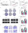
HDAC2 overexpression partly recovered the inhibitory effects of miR-490-5p. (A) Transfection efficiency in OS cells was confirmed by western blot analysis. (B,C) The CCK-8 assay indicated that the inhibitory effects of miR-490-5p overexpression in OS cells were substantially reduced by HDAC2 overexpression. (D,E) Overexpression of HDAC2 partly reversed the suppressed effects of miR-490-5p overexpression on the colony formation capability of U-2OS and 143B cells (crystal violet staining). (F,G) The wound healing assay revealed that HDAC2 overexpression reversed miR-490-5p-mediated inhibition of OS cell migration. Scale bar =100 µm. (H,I) The transwell invasion assay showed that HDAC2 overexpression partially restored the cell invasion inhibition mediated by miR-490-5p (crystal violet staining). Scale bar =50 µm. The results are presented as the mean ± SD. ***, P<0.001. CCK-8, Cell Counting Kit-8; OS, osteosarcoma; SD, standard deviation.

Similar articles

References

    1. Zuo D, Shogren KL, Zang J, et al. Inhibition of STAT3 blocks protein synthesis and tumor metastasis in osteosarcoma cells. J Exp Clin Cancer Res 2018;37:244 . 10.1186/s13046-018-0914-0 - DOI - PMC - PubMed
    1. Sayles LC, Breese MR, Koehne AL, et al. Genome-Informed Targeted Therapy for Osteosarcoma. Cancer Discov 2019;9:46-63. 10.1158/2159-8290.CD-17-1152 - DOI - PMC - PubMed
    1. Ma H, He C, Cheng Y, et al. PLK1shRNA and doxorubicin co-loaded thermosensitive PLGA-PEG-PLGA hydrogels for osteosarcoma treatment. Biomaterials 2014;35:8723-34. 10.1016/j.biomaterials.2014.06.045 - DOI - PubMed
    1. Whelan JS, Jinks RC, McTiernan A, et al. Survival from high-grade localised extremity osteosarcoma: combined results and prognostic factors from three European Osteosarcoma Intergroup randomised controlled trials. Ann Oncol 2012;23:1607-16. 10.1093/annonc/mdr491 - DOI - PMC - PubMed
    1. Wang X, Qin G, Liang X, et al. Targeting the CK1α/CBX4 axis for metastasis in osteosarcoma. Nat Commun 2020;11:1141 . 10.1038/s41467-020-14870-4 - DOI - PMC - PubMed

LinkOut - more resources