

En-Face OCT and Microperimetric Analysis of Intraretinal Microcysts in Eyes After Pars Plana Vitrectomy for Epiretinal Membrane
- PMID: 40792187
- PMCID: PMC12338324
- DOI: 10.2147/OPTH.S524057
En-Face OCT and Microperimetric Analysis of Intraretinal Microcysts in Eyes After Pars Plana Vitrectomy for Epiretinal Membrane
Abstract
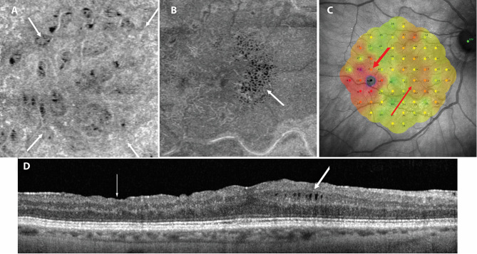
Purpose: To compare the location of microcystic macular edema (MME) with areas of retinal nerve fiber layer (RNFL) damage in the macula detected on en-face SDOCT in eyes that underwent pars plana vitrectomy (PPV) due to the epiretinal membrane (ERM).
Patients and methods: Thirty-five eyes were enrolled at least 6 months after PPV with removal of ERM and inner limiting membrane (ILM). In each eye, en-face SDOCT and microperimetry were performed. The area of RNFL damage was measured and compared with the position of MME and correlated with the volume of retinal layers and retinal sensitivity (AT).
Results: MME was observed in 17 eyes (48.6%) in the area devoid of ILM, often in places with arcuate damage to the RNFL bundle. The mean area of RNFL damage in eyes with MME was 9.03 ± 5.3 mm2 and was significantly larger than in eyes where microcysts were not present, where it measured 3.92 ± 3.3 mm2. A significant negative correlation was observed between the area of RNFL damage and GCL volume and AT.
Conclusion: The topographic analysis of the MME position in eyes after PPV due to ERM confirmed the association of this pathology with ganglion cells and RNFL damage related to the removal of the ILM and ERM. There are probably two pathways leading to the development of MME: one starting from Muller cell damage during ILM peeling and the other due to retrograde death of ganglion cells in the areas of arcuate RNFL defects.
Keywords: en-face OCT; epiretinal membrane; ganglion cell layer; microcystic macular edema; microperimetry; retinal nerve fiber layer.
© 2025 Kaluzny et al.
Conflict of interest statement
The authors have not received grant support or research funding, and they do not have any proprietary interests in the materials described in the article.
Figures

References
LinkOut - more resources
Full Text Sources
Research Materials
Miscellaneous
