

A Case of Pyogenic Vertebral Osteomyelitis and Iliopsoas Abscess Caused by Invasive Pneumococcal Disease Serotype 35F: Utility of Diffusion-Weighted Whole-Body Imaging With Background Body Signal Suppression as an Adjunctive Diagnostic Tool
- PMID: 40792325
- PMCID: PMC12337593
- DOI: 10.7759/cureus.87770
A Case of Pyogenic Vertebral Osteomyelitis and Iliopsoas Abscess Caused by Invasive Pneumococcal Disease Serotype 35F: Utility of Diffusion-Weighted Whole-Body Imaging With Background Body Signal Suppression as an Adjunctive Diagnostic Tool
Abstract
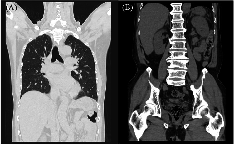
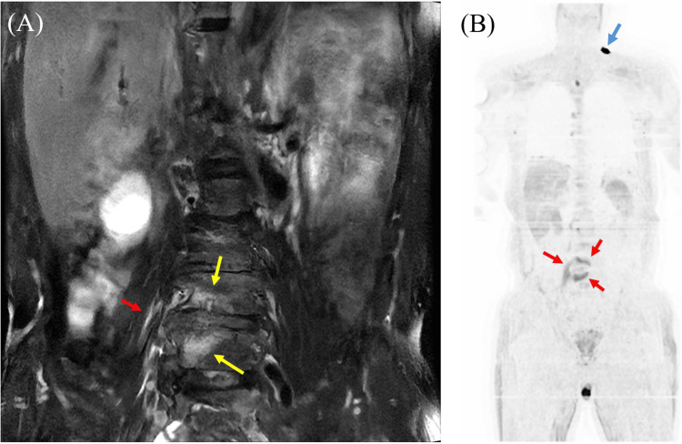
We present a case of invasive pneumococcal disease (IPD) complicated by both iliopsoas abscess and pyogenic vertebral osteomyelitis caused by serotype 35F Streptococcus pneumoniae in a 64-year-old man with a history of splenectomy who was unvaccinated. The patient experienced difficulty moving, severe back pain, vomiting, and high fever. Advanced imaging techniques, including T2-weighted lumbar MRI and diffusion-weighted whole-body imaging with background body signal suppression (DWIBS), revealed hyperintensities in the left iliopsoas muscle and L4-L5 vertebral bodies, facilitating diagnosis. Blood cultures confirmed the presence of serotype 35F S. pneumoniae, a non-vaccine type associated with an increased risk of invasive disease and mortality. The patient was successfully treated with targeted antibiotics and disc lavage, resulting in symptom resolution. To our knowledge, this is the first reported case of serotype 35F S. pneumoniae causing both iliopsoas abscess and vertebral osteomyelitis, making it a noteworthy contribution to medical literature. Notably, DWIBS proved to be a valuable adjunct diagnostic tool, highlighting its potential for visually accessible, comprehensive screening of inflammation and abscesses throughout the body. We believe that DWIBS is particularly useful when bacteria capable of inducing lesions in multiple organs, such as S. pneumoniae or Staphylococcus aureus, are isolated from blood cultures. Although DWIBS is still emerging in infectious disease diagnostics, this case underscores its promising role in detecting abscesses and inflammatory lesions.
Keywords: diffusion-weighted imaging (dwi); dwibs; iliopsoas abscess; invasive pneumococcal disease; mri images; pyogenic vertebral osteomyelitis; streptococcus pneumoniae.
Copyright © 2025, Kimura et al.
Conflict of interest statement
Human subjects: Informed consent for treatment and open access publication was obtained or waived by all participants in this study. Conflicts of interest: In compliance with the ICMJE uniform disclosure form, all authors declare the following: Payment/services info: All authors have declared that no financial support was received from any organization for the submitted work. Financial relationships: All authors have declared that they have no financial relationships at present or within the previous three years with any organizations that might have an interest in the submitted work. Other relationships: All authors have declared that there are no other relationships or activities that could appear to have influenced the submitted work.
Figures
Similar articles
-
Immunogenicity and seroefficacy of pneumococcal conjugate vaccines: a systematic review and network meta-analysis.Health Technol Assess. 2024 Jul;28(34):1-109. doi: 10.3310/YWHA3079. Health Technol Assess. 2024. PMID: 39046101 Free PMC article.
-
A case of infectious endocarditis and vertebral discitis caused by Streptococcus pneumoniae serotype 23A.J Infect Chemother. 2025 Aug;31(8):102749. doi: 10.1016/j.jiac.2025.102749. Epub 2025 Jun 7. J Infect Chemother. 2025. PMID: 40490099
-
Signs and symptoms to determine if a patient presenting in primary care or hospital outpatient settings has COVID-19.Cochrane Database Syst Rev. 2022 May 20;5(5):CD013665. doi: 10.1002/14651858.CD013665.pub3. Cochrane Database Syst Rev. 2022. PMID: 35593186 Free PMC article.
-
An Elderly Patient With Lumbar Spondylodiscitis and Psoas Abscess: Diagnostic Considerations in the Context of Aerococcus urinae.Cureus. 2025 May 24;17(5):e84752. doi: 10.7759/cureus.84752. eCollection 2025 May. Cureus. 2025. PMID: 40551937 Free PMC article.
-
Sickle Cell Disease.2003 Sep 15 [updated 2025 Feb 13]. In: Adam MP, Feldman J, Mirzaa GM, Pagon RA, Wallace SE, Amemiya A, editors. GeneReviews® [Internet]. Seattle (WA): University of Washington, Seattle; 1993–2025. 2003 Sep 15 [updated 2025 Feb 13]. In: Adam MP, Feldman J, Mirzaa GM, Pagon RA, Wallace SE, Amemiya A, editors. GeneReviews® [Internet]. Seattle (WA): University of Washington, Seattle; 1993–2025. PMID: 20301551 Free Books & Documents. Review.
References
-
- The burden of pneumococcal disease among adults in developed and developing countries: what is and is not known. Fedson DS, Scott JA. Vaccine. 1999;17:11–18. - PubMed
-
- The role of Streptococcus pneumoniae virulence factors in host respiratory colonization and disease. Kadioglu A, Weiser JN, Paton JC, Andrew PW. Nat Rev Microbiol. 2008;6:288–301. - PubMed
Publication types
LinkOut - more resources
Full Text Sources