Review
. 2025 Dec;14(1):2544726.
doi: 10.1080/22221751.2025.2544726. Epub 2025 Aug 17.

Regulation of viral hepatitis by N6-methyladenosine RNA methylation

Affiliations
Review

Regulation of viral hepatitis by N6-methyladenosine RNA methylation

Jae-Su Moon et al. Emerg Microbes Infect. 2025 Dec.

Abstract

Recent studies have shown that the presence of an RNA modification, N6-methyladenosine (m6A), in viral RNAs during infection significantly impacts the outcome of viral replication and pathogenesis. In particular, various functions of m6A have been elucidated in hepatitis B virus (HBV), hepatitis C virus (HCV), and hepatitis delta virus (HDV). During viral infection, m6A methylation not only directly affects the replication of these viruses but also regulates diverse cellular RNAs to control pathogenesis. This review aims to explore the functions of m6A modification in the infectious processes and pathogenesis of HBV, HCV, and HDV. Understanding the role of m6A methylation in these viral life cycles is essential for elucidating their pathogenesis and developing novel therapeutic strategies for HBV, HCV, and HDV infections.

Keywords: Hepatitis B virus; N6-methyladenosine RNA methylation; hepatitis C virus; hepatitis Delta virus; viral hepatitis.


Conflict of interest statement

No potential conflict of interest was reported by the author(s).

Figures
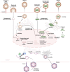
Figure 1.
Figure 1.
Overview of the life cycles of HBV, HCV, and HDV. HBV life cycle: HBV virions infect human hepatocytes by binding to the sodium taurocholate co-transporting polypeptide (NTCP) receptor, mediated by the epidermal growth factor receptor (EGFR) as a co-receptor. After infection, the HBV nucleocapsid is transported to the nuclear membrane, releasing its relaxed circular DNA (rcDNA) into the nucleus. Within the nucleus, the rcDNA genome is converted into cccDNA by viral polymerase and host factors. The cccDNA is highly resistant to host antiviral responses and can persist in hepatocytes for long periods, contributing to chronic infection. HBV cccDNA encodes the viral proteins: surface (HBs), pre-core or e (HBe), and core (HBc) antigen, polymerase, and X (HBx). The viral polymerase interacts with pgRNA to initiate nucleocapsid assembly, and pgRNA serves as a template to produce rcDNA within nucleocapsid by reverse transcription. The mature HBV capsid either re-enters the nucleus to maintain cccDNA or is secreted from the cell, acquiring an envelope composed of viral envelope proteins embedded in the host cell lipid membrane during the process. HCV life cycle: The infection of HCV into hepatocytes is mediated by several receptors, including CD81, scavenger receptor class B type (SR-BI), and claudin-1. After attachment, HCV virions enter the cell via clathrin-mediated endocytosis. HCV RNA is then directly translated into a single polyprotein precursor, which is cleaved into three structural and seven nonstructural proteins by viral and host proteases. The non-structural proteins, including NS3/4A, NS5A, and NS5B, are involved in viral replication. HCV RNA replication occurs in membrane-associated complexes derived from the ER, known as the membranous web. The viral polymerase, NS5B, possesses RNA-dependent RNA polymerase activity, enabling it to synthesize negative-sense viral RNA using a positive-sense HCV RNA template. Subsequently, a positive-sense HCV RNA is synthesized from a negative-sense RNA by NS5B, and a new viral RNA genome is packaged into the virions. HDV life cycle: HDV, a defective virus, relies on the HBV life cycle for its propagation. HDV uses the envelope proteins of HBV to egress from and re-enter hepatocytes, indicating that HDV enters the hepatocytes through the same NTCP receptor as HBV. After infection, HDV releases its ribonucleoprotein into the cytoplasm, from where it is transported to the nucleus. HDV replicates using RNA-directed RNA synthesis, producing an anti-genome entirely complementary to the HDV genome, but does not translate antigens. This anti-genome is used as a template to produce the HDV genome. This replication occurs via host RNA polymerase II through a rolling-circle mechanism facilitated by self-cleaving RNA sequences (ribozymes). HDV mRNAs are synthesized from the HDV genome and translated at the ER to produce HDAg proteins, which then re-enter the nucleus to enhance genome replication. Small and large HDAg (S-HDAg and L-HDAg) proteins associate with newly synthesized genomic RNA to form new RNPs. These RNPs are transported back to the cytoplasm, where L-HDAg aids in linking to HBV surface antigen (HBsAg) in the ER, leading to the assembly of new viral particles. These viral particles are then released from hepatocytes via the Golgi apparatus to infect adjacent cells.
Figure 2.
Figure 2.
Cellular m6A machinery: m6A methyltransferases, m6A demethylases, and m6A reader proteins. m6A RNA methylation of the adenosine base at the nitrogen 6 position is the most abundant and well-characterized modification of cellular RNAs. This modification is predominantly enriched in the 5’ and 3'-untranslated regions (UTRs) and adjacent to the stop codon. m6A methylation is reversibly catalyzed by m6A “writers” (the METTL3, METTL14, and WTAP complex) and removed by “erasers” (FTO or ALKBH5). For the regulation of m6A methylation-mediated RNA function, m6A “reader” proteins must bind to m6A methylated RNA, and then these proteins regulate m6A-methylated RNA stability, turnover, and translation.
Figure 3.
Figure 3.
The roles of m6A methylation in modulating the HBV life cycle. m6A methylation occurs at 1907A, located in the lower stem of the epsilon element. HBV pgRNA includes the epsilon element at both 5’ and 3’ ends due to terminal redundancy, but the other HBV RNAs contain this motif only ate the 3’ end of their viral RNAs. m6A methylation at the 5’ epsilon of pgRNA enhances the nucleocapsid assembly by upregulating its interaction with the core. Additionally, this methylation recruits YTHDF2 and 3 inhibits RIG-I sensing to suppress the immune response. Conversely, m6A methylation at the 3’ epsilon of HBV RNAs decreases RNA stability by the recruitment of YTHDF2. Moreover, m6A modifications at both the 3’ and 5’ epsilon regions facilitate increased nuclear export of the RNA through interactions with YTHDC1 and FMRP. In the context of host genes, m6A methylation of PTEN mRNA is increased during HBV infection, and m6A methylation of the 3’ UTR of PTEN mRNA decreases its stability via YTHDF2 binding. Reduced PTEN expression attenuates the immune response by inhibiting IRF3 nuclear localization and promotes hepatocarcinogenesis via activation of PI3 K/AKT signalling pathway. HBx enhances nuclear import of METTL3/14 complex and recruits these methyltransferases into HBV cccDNA and the PTEN chromosome locus to add m6A RNA methylation.
Figure 4.
Figure 4.
The roles of m6A methylation in modulating the HCV life cycle. m6A methylation occurs at multiple sites across the HCV genome and plays a significant role in the viral life cycle. Specifically, m6A methylation within the IRES region increases IRES-dependent translation by recruiting YTHDC2. In addition, m6A methylation in E1 coding region decreases virion assembly by the down-regulation of interaction between the HCV genome and core, while methylation in the NS5B coding region suppresses innate immune response via the abolishing of the viral genome sensing by RIG-I. During HCV infection, alterations in m6A methylation also occur in host mRNAs, affecting various cellular processes. These changes in cellular signalling pathways can lead to either an increase or a decrease in HCV replication and contribute to the development of HCC.
Figure 5.
Figure 5.
The roles of m6A methylation in the regulation of the HDV life cycle. m6A methylation in the HDV genome and its impact on virion assembly. m6A methylation of the HDV genome recruits YTHDF1. YTHDF1 binding to m6A methylated HDV RNA interferes with HDAg binding, thereby reducing RNP formation and virion assembly. Specific m6A sites in HDV are not yet identified; the illustration depicts the general effect of m6A on HDV RNP formation.

References

    1. Seeger C, Mason WS.. Hepatitis B virus biology. Microbiol Mol Biol Rev. 2000;64(1):51–68. doi: 10.1128/MMBR.64.1.51-68.2000 - DOI - PMC - PubMed
    1. Paul D, Madan V, Bartenschlager R.. Hepatitis C virus RNA replication and assembly: living on the fat of the land. Cell Host Microbe. 2014;16(5):569–579. doi: 10.1016/j.chom.2014.10.008 - DOI - PMC - PubMed
    1. Wedemeyer H, Negro F.. Devil hepatitis D: an orphan disease or largely underdiagnosed? Gut. 2019;68(3):381–382. doi: 10.1136/gutjnl-2018-317403 - DOI - PubMed
    1. Hu J, Protzer U, Siddiqui A.. Revisiting hepatitis B virus: challenges of curative therapies. J Virol. 2019;93(20): e01032–19. - PMC - PubMed
    1. Manns MP, Maasoumy B.. Breakthroughs in hepatitis C research: from discovery to cure. Nat Rev Gastroenterol Hepatol. 2022;19(8):533–550. doi: 10.1038/s41575-022-00608-8 - DOI - PMC - PubMed

MeSH terms

LinkOut - more resources