. 2025 Aug;29(15):e70773.
doi: 10.1111/jcmm.70773.

The Transcription Factor CREB1 Triggers the Progression of Clear Cell Renal Cell Carcinoma by Promoting CENPE Expression

Affiliations

The Transcription Factor CREB1 Triggers the Progression of Clear Cell Renal Cell Carcinoma by Promoting CENPE Expression

Hao Jiang et al. J Cell Mol Med. 2025 Aug.

Abstract

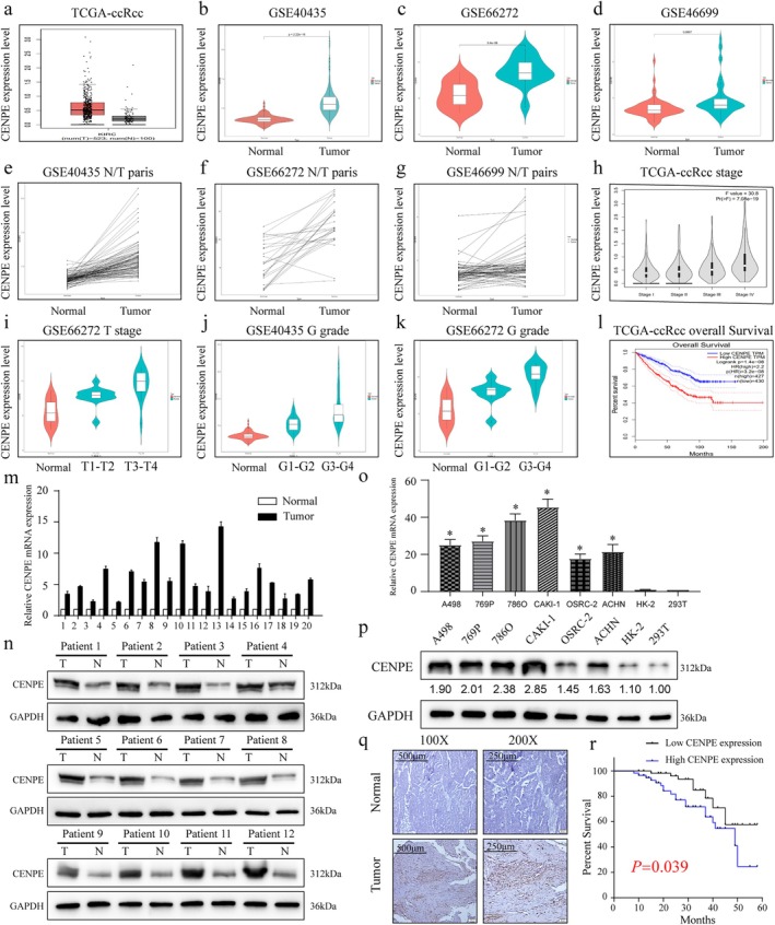
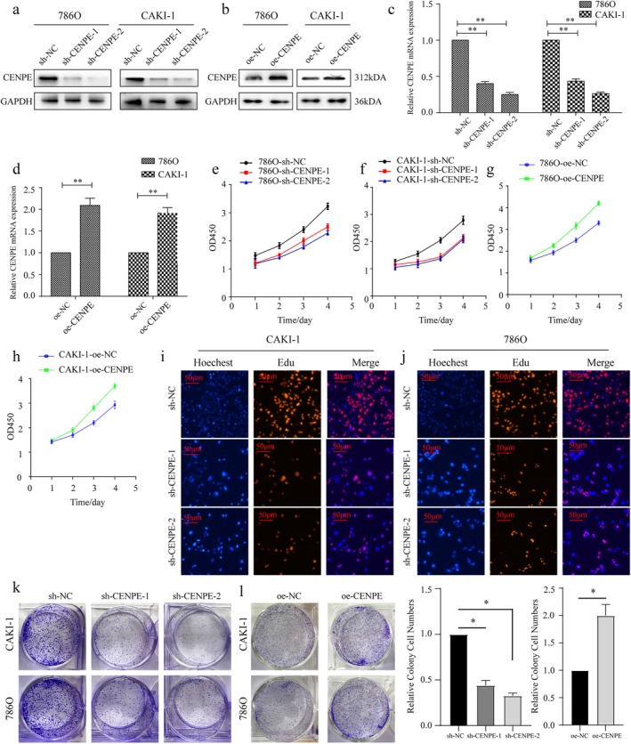
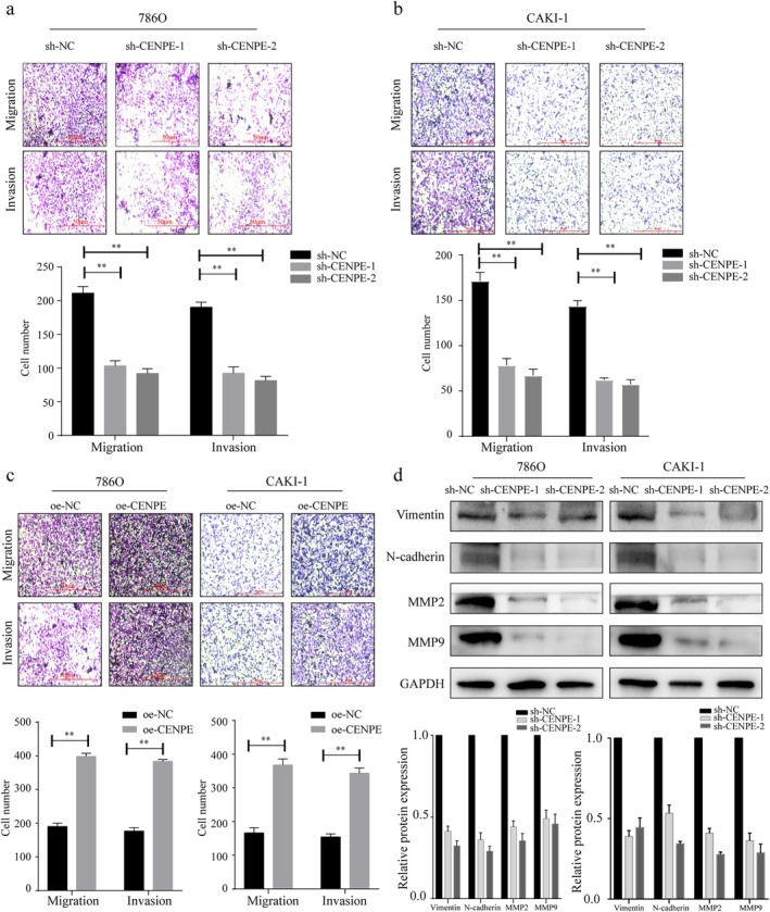
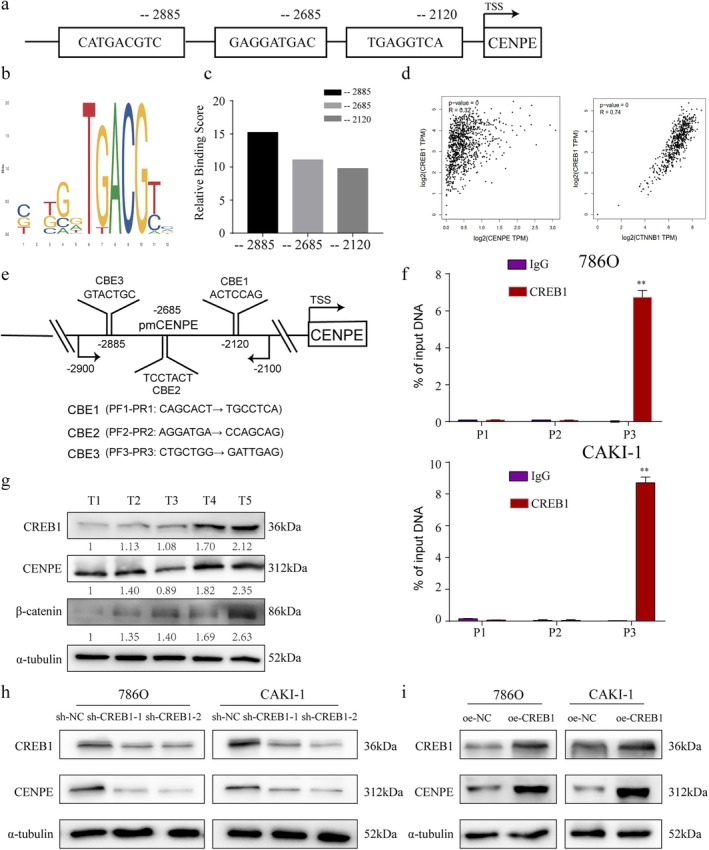
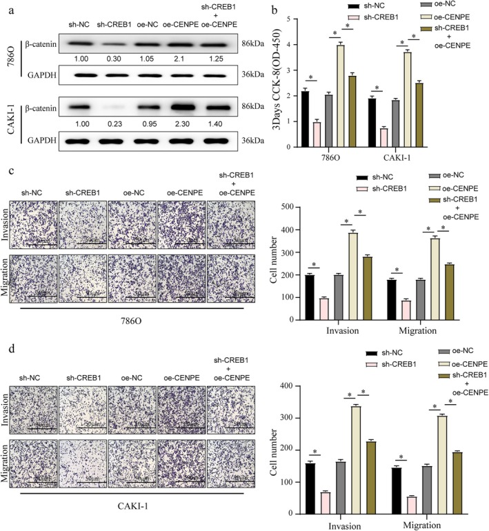
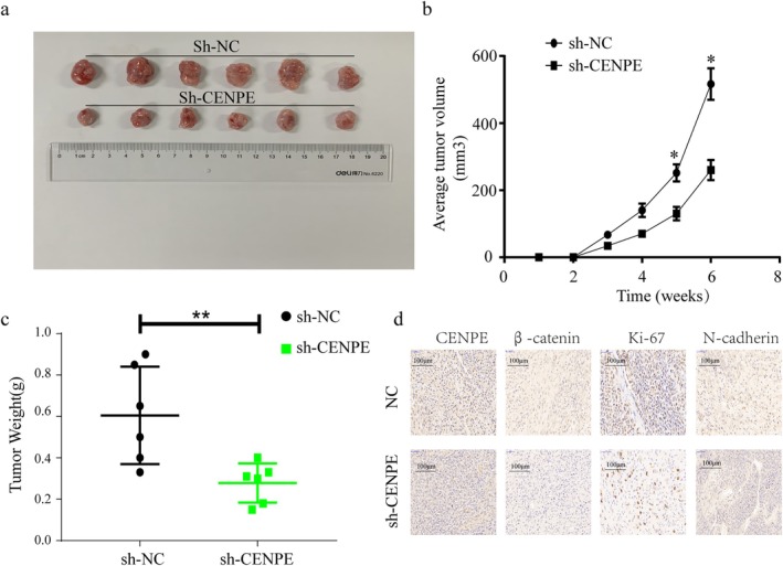
Centromere-associated protein E (CENPE) has been identified as overexpressed in multiple cancers and exerts a tumour promotion function by affecting chromosome misalignment and mitosis. However, the expression pattern, biological roles, and underlying molecular mechanism of CENPE in clear cell renal cell carcinoma (ccRCC) progression have not been fully elucidated. In the present study, the expression levels of CENPE in ccRCC and paracancerous specimens were measured using the public RNA sequencing data and validated in a cohort of ccRCC samples from our centre. We found that CENPE was significantly over-expressed in ccRCC tissues and promoted proliferative and metastatic abilities of ccRCC cells and xenografts through regulating the epithelial-mesenchymal transition (EMT) process. Furthermore, bioinformatic analysis and ChIP assay indicated that the transcription factor CREB1 bound to the promoter region of CENPE and activated its transcription in ccRCC cells. Taken together, our findings demonstrated that the CREB1-CENPE axis was responsible for stimulating the in vitro and in vivo progression of ccRCC, serving as a promising therapeutic target for ccRCC.

Keywords: CENPE; CREB1; EMT; ccRCC; progression.


Conflict of interest statement

The authors declare no conflicts of interest.

Figures

FIGURE 1
FIGURE 1
Overexpression of CENPE in ccRCC. (a) Expression levels of CENPE in the TCGA‐KIRC dataset containing 523 kidney renal clear cell carcinoma specimens and 100 normal specimens (Red: Tumour; Grey: Normal). (b–d) Expression levels of CENPE in ccRCC specimens and adjacent non‐tumoral renal specimens of the GSE40435 (b), GSE66272 (c) and GSE46699 datasets (d). (e–g) Pairwise expression levels of CENPE in ccRCC and adjacent non‐tumoral specimens of the GSE40435 (e), GSE66272 (f) and GSE46699 datasets (g). (h) Violin plots visualising CENPE levels in ccRCC specimens at stage I‐IV. (i, k) Expression levels of CENPE in ccRCC specimens classified by T staging (i) and G staging (k) using the GSE66272 dataset. (j) Expression levels of CENPE in ccRCC specimens classified by G staging using the GSE40435 dataset. (l) Overall survival of ccRCC patients stratified by the expression level of CENPE (HR, Hazard Ratio). (m, n) Expression levels of CENPE in ccRCC specimens and paired non‐tumoral specimens collected in our centre. (o, p) Expression levels of CENPE in ccRCC cell lines and normal cell lines, the numbers below represent the relative CENPE expression normalised to GAPDH, with 293 T cells set as the reference (1.00). (q) Immunohistochemical staining of CENPE in ccRCC specimens and paired non‐tumoral specimens collected in our centre (magnification = 100× and 200×), with brown staining indicates positive expression of CENPE, while blue staining (haematoxylin) marks the cell nuclei. (r) Kaplan–Meier survival of ccRCC patients classified by the median level of CENPE. *p < 0.05.
FIGURE 2
FIGURE 2
CENPE promotes the proliferation of ccRCC in vitro. (a, b) Transfection efficacy of sh‐CENPE‐1, sh‐CENPE‐2 (a) and oe‐CENPE (b) in 786O and CAKI‐1 cells detected by Western blot. (c, d) Transfection efficacy of sh‐CENPE‐1, sh‐CENPE‐2 (c) and oe‐CENPE (d) in 786O and CAKI‐1 cells detected by qRT‐PCR. (e, f) Cell viability in 786O (e) and CAKI‐1 cells (f) transfected with sh‐CENPE‐1 or sh‐CENPE‐2 detected by CCK‐8 assay. (g, h) Cell viability in 786O (g) and CAKI‐1 cells (h) transfected with oe‐CENPE detected by CCK‐8 assay. (i, j) EdU‐positive cells (red) and Hoechest‐staining nuclei (blue) in CAKI‐1 (i) and 786O cells (j) transfected with sh‐CENPE‐1 or sh‐CENPE‐2 (scale bar = 50 μm). (k, l) Colony formation in CAKI‐1 and 786O cells transfected with sh‐CENPE‐1 or sh‐CENPE‐2 (k) and oe‐CENPE (l). (m) Bar plots show the relative colony numbers in 786O cells following CENPE knockdown or overexpression. *p < 0.05, **p < 0.01.
FIGURE 3
FIGURE 3
CENPE promotes the metastasis of ccRCC in vitro. (a, b) Migration and invasion assays in 786O (a) and CAKI‐1 cells (b) transfected with sh‐CENPE‐1 or sh‐CENPE‐2 (scale bar = 50 μm). (c) Migration and invasion assays in 786O and CAKI‐1 cells transfected with oe‐CENPE (scale bar = 50 μm). (d) Protein expressions of Vimentin, N‐cadherin, MMP2 and MMP9 in 786O and CAKI‐1 cells transfected with sh‐CENPE‐1 or sh‐CENPE‐2, analysed by Western blot. **p < 0.01.
FIGURE 4
FIGURE 4
CREB1 activates the transcription of CENPE in ccRCC. (a, b) CREB1 binding sites in the promoter region of CENPE predicted by the JASPER database (a) and the binding region within the predicted binding site (b). (c) Relative score for each potential binding sequence of CENPE combined with CREB1. (d) Correlations of CREB1 with CENPE and CTNNB1 (β‐catenin) in the TCGA dataset. (e) Schematic of the CENPE promoter region showing the designed primers (CBE1, CBE2, CBE3) for ChIP assay. (f) ChIP assay visualising the input of CREB1 and IgG in the promoter region of CENPE (−2885). (g) Protein expressions of CREB1, CENPE and β‐catenin in ccRCC specimens. (h) Protein expressions of CREB1 and CENPE in 786O and CAKI‐1 cells transfected with sh‐CENPE‐1 or sh‐CENPE‐2. (i) Protein expressions of CREB1 and CENPE in 786O and CAKI‐1 cells transfected with oe‐CREB1. **p < 0.01.
FIGURE 5
FIGURE 5
CREB1 is essential for the oncogenic role of CENPE in ccRCC. Protein expression of β‐catenin (a), cell viability (b), migration (c) and invasion (d) in 786O and CAKI‐1 cells treated with blank control, or transfected with sh‐CREB1, oe‐CENPE or sh‐CREB1 + oe‐CENPE. Scale bar = 50 μm. *p < 0.05.
FIGURE 6
FIGURE 6
The CREB1‐CENPE axis favours in vivo growth of ccRCC. Five‐week‐old female nude mice were subcutaneously injected with 7 × 106 CAKI‐1 cells transfected with sh‐NC (n = 6) and sh‐CENPE (n = 6) into the posterior flank. (a) Representative images of xenograft tumours derived from CAKI‐1 cells transfected with sh‐NC (control) or sh‐CENPE after 6 weeks of growth. (b) Average tumour volume measured at indicated time points. (c) Tumour weight of excised xenografts at the end of the experiment. (d) Immunohistochemical staining of CENPE, β‐catenin, Ki‐67 and N‐cadherin in ccRCC xenografts. n = 6. Scale bar = 50 μm. *p < 0.05, **p < 0.01.

Similar articles

References

    1. Siegel R. L., Giaquinto A. N., and Jemal A., “Cancer Statistics, 2024,” CA: A Cancer Journal for Clinicians 74, no. 1 (2024): 12–49. - PubMed
    1. Gao B., Wang L., Zhang Y., et al., “MiR‐532‐3p Suppresses Cell Viability, Migration and Invasion of Clear Cell Renal Cell Carcinoma Through Targeting TROAP,” Cell Cycle 20, no. 16 (2021): 1578–1588. - PMC - PubMed
    1. Liu W., Zhang B., Zhang D., et al., “The RBPJ/DAPK3/UBE3A Signaling Axis Induces PBRM1 Degradation to Modulate the Sensitivity of Renal Cell Carcinoma to CDK4/6 Inhibitors,” Cell Death & Disease 13, no. 4 (2022): 295. - PMC - PubMed
    1. Jonasch E., Walker C. L., and Rathmell W. K., “Clear Cell Renal Cell Carcinoma Ontogeny and Mechanisms of Lethality,” Nature Reviews. Nephrology 17, no. 4 (2021): 245–261. - PMC - PubMed
    1. El‐Arabey A. A., Salama S. A., and Abd‐Allah A. R., “CENP‐E as a Target for Cancer Therapy: Where Are We Now?,” Life Sciences 208 (2018): 192–200. - PubMed

MeSH terms

Substances

LinkOut - more resources