. 2025 Aug 12;21(8):e1013427.
doi: 10.1371/journal.ppat.1013427. eCollection 2025 Aug.

Transcriptome profiling of L. infantum-infected human macrophages reveals sex-specific type I interferon induction

Affiliations

Transcriptome profiling of L. infantum-infected human macrophages reveals sex-specific type I interferon induction

Annika Bea et al. PLoS Pathog. .

Abstract

Sex-based differences in the immune system influence the clinical course of infectious diseases, including many parasitic infections. Field studies of human infections and controlled experimental rodent models have shown that certain clinical forms of leishmaniasis occur more frequently in males. Leishmania parasites infect and proliferate in innate immune cells, particularly macrophages, and modulate early immune responses that constrain their survival and replication. In this study, we used a high-throughput in vitro system to assess sex differences in human macrophage-specific immunity to Leishmania (L.) infantum infection. Quantification of infection showed significantly higher infection rates and parasite loads in macrophages derived from men compared to those from women up to 76 hours post-infection (hpi). Evaluation of the macrophage phenotype during L. infantum infection revealed only minor changes in the proportions of primarily proinflammatory M1-like macrophages, whereas a reduction in the anti-inflammatory M2-like phenotype was observed in both sexes. Cytokine profiling revealed elevated levels of TNF, IL-8, IL-10, and reduced levels of IL-18 and CCL2 in culture supernatants over the time of infection. Transcriptomic analysis showed the highest adaptation of gene expression at 6 hpi, which was more pronounced in female-derived macrophages (1428 down-regulated/2145 up-regulated genes) compared to male-derived macrophages (972 down-regulated/1637 up-regulated genes), and gradually decreased over time in both sexes. Genes associated with type I interferon responses (e.g., IFIT2, IFIT3, IFIT5, OASL, JAK1), specific cytokine response (IL-15, IL-1R1), and the matrix metalloproteinase MMP9 were up-regulated in female macrophages, while genes encoding proinflammatory chemokines involved in immune cell recruitment (CXCL1, CXCL3, CCL20, CCL7) were up-regulated in male macrophages. Treatment of infected macrophages with estradiol conferred marginal resistance to infection in female-derived macrophages, whereas testosterone treatment had no effect. In summary, our findings reveal immune mediators and underscore a biological sex difference that may explain females' superior ability to combat Leishmania infections.


Conflict of interest statement

The authors have declared that no competing interests exist.

Figures
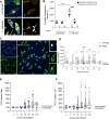
Fig 1
Fig 1. Intracellular localization of Leishmania infantum and sex-specific differences in human macrophage infection.
Fig 2
Fig 2. L. infantum infection induces changes in M1/M2 polarization of human macrophages.
Fig 3
Fig 3. Infection-specific cytokines produced during L. infantum-infection in macrophages derived from men and women.
Mature macrophages from female and male donors were infected with L. infantum (MOI 15:1) for 6 to 52 hours. Cytokines were determined in the culture supernatant using cytometric bead assay (LEGENDplex). Time-course analysis of the MFI of (A) TNF (B) IL-8 (C) IL-10 (D) CCL2 and (E) IL-18 at indicated time points (left side) and respective area under the curve (AUC) (right side). Data represent mean ± SEM, nF/M = 4/4. P-values were calculated using two-tailed paired analysis Student´s t-test (*p < 0.05, **p < 0.01).
Fig 4
Fig 4. Analysis of differentially expressed genes between uninfected and infected macrophages from female and male donors.
Mature macrophages from women and men were infected with L. infantum (MOI 15:1). mRNA from infected macrophages and naïve controls was isolated and subjected to whole transcriptome sequencing. Number of differentially expressed genes (adj. p < 0.05) at different time points after L. infantum infection in relation to uninfected controls for macrophages from (A) women and (B) men. Box length depicts number of differently expressed genes down-regulated (left, blue) and up-regulated (right, red). Color shading correlates with the magnitude of regulation (dark: log2(fold change)>3; medium: 1 < log2(fold change) <3, light: log2(fold change) <1). (C, D) Venn diagrams depicting differentially regulated genes (adj. p < 0.05) at different time points after L. infantum infection of macrophages from (C) women and (D) men compared to uninfected controls. nF/M = 4/4.
Fig 5
Fig 5. Transcriptional analysis of L. infantum-infected macrophages from female and male donors 6 hpi.
Mature macrophages from (A-D) women and (E-H) men were infected with L. infantum (MOI 15:1) for 6 hours. mRNA from infected macrophages and naïve controls was isolated and subjected to whole transcriptome sequencing. (A, E) Volcano plots depicting differential gene expression between uninfected and infected macrophages according to magnitude of change (Log2(fold change)) and statistical significance of change (-Log10(adjusted p-value). Genes significantly regulated between the conditions (Log2(fold change) > I0.25I, adj. p < 0.05) are marked in red (left side = low in infection, right side = high in infection). (B, F) Heatmaps depict top 30 regulated genes. (D, H) KEGG pathway analysis of significantly up-regulated genes in infection (adj. p < 0.05, log2 (fold change) >1) and (C, G) Biological processes GO term enrichment of significantly down-regulated genes in infection (adj. p < 0.05, log2 (fold change) <-1). (C, D, G, H) Shown are the top 20 pathways/processes sorted by fold enrichment. Significance of enrichment indicated by color and number of genes in pathway indicated in node size. nF/M = 4/4.
Fig 6
Fig 6. Sex differences in the transcriptome of L. infantum-infected macrophages.
Mature macrophages from female and male donors were infected with L. infantum (MOI 15:1) for 6 hours. mRNA from infected macrophages and naïve controls was isolated and subjected to whole transcriptome sequencing. (A-C) Venn diagrams depicting the DEGs between female and male donors following L. infantum infection compared to corresponding naïve control (adj. p < 0.05, log2 (fold change) <|1|) at (A) 6, (B) 16 and (C) 52 hpi. For 6 hpi (D-I) top enriched pathways identified using Reactome database are depicted at the side of the heatmap. Heatmaps depict selected corresponding genes for (D) exclusively down-regulated genes in female samples, (E) similarly down-regulated genes in both female and male samples and (F) exclusively down-regulated genes in male samples, (G) exclusively up-regulated genes in female samples, (H) similarly up-regulated genes in both female and male samples and (I) exclusively up-regulated genes in male samples,.
Fig 7
Fig 7. Influence of steroid hormone stimulation on L. infantum infection of macrophages.
Mature macrophages from female and male donors were stimulated with 17-Estradiol (E2) in various concentrations (0.01– 5μM) before infection with L. infantum (MOI 15:1) for 28 hours. Parameters were quantified using Opera Phenix confocal microscope and customized image analysis sequence. (A) Infection rate and (B) parasite burden of macrophages normalized to corresponding infected unstimulated control (POC = 100). Data are shown as boxplot, E2 = nF/M - = 12/13, nF/M 0.01µM = 11/12, nF/M 0.1µmM = 5/7, nF/M 0.5µM = 6/8, nF/M 1µM = 6/8, nF/M 2µM = 6/8, nF/M 5µM = 5/7.

References

    1. Klein SL, Flanagan KL. Sex differences in immune responses. Nat Rev Immunol. 2016;16(10):626–38. - PubMed
    1. Sellau J, Groneberg M, Lotter H. Androgen-dependent immune modulation in parasitic infection. Semin Immunopathol. 2019;41(2):213–24. doi: 10.1007/s00281-018-0722-9 - DOI - PubMed
    1. Murray HW, Berman JD, Davies CR, Saravia NG. Advances in leishmaniasis. Lancet. 2005;366(9496):1561–77. doi: 10.1016/S0140-6736(05)67629-5 - DOI - PubMed
    1. Biswas SK, Chittezhath M, Shalova IN, Lim J-Y. Macrophage polarization and plasticity in health and disease. Immunol Res. 2012;53(1–3):11–24. doi: 10.1007/s12026-012-8291-9 - DOI - PubMed
    1. Tomiotto-Pellissier F, Bortoleti BT da S, Assolini JP, Gonçalves MD, Carloto ACM, Miranda-Sapla MM, et al. Macrophage Polarization in Leishmaniasis: Broadening Horizons. Front Immunol. 2018;9:2529. doi: 10.3389/fimmu.2018.02529 - DOI - PMC - PubMed