. 2025 Aug 12;15(1):29546.
doi: 10.1038/s41598-025-13617-9.

Pathology of stranded blue sharks (Prionace glauca) impaled by swordfish (Xiphias gladius)

Affiliations

Pathology of stranded blue sharks (Prionace glauca) impaled by swordfish (Xiphias gladius)

A Campos-Sosa et al. Sci Rep. .

Abstract

This article describes the pathological findings of ten blue sharks (Prionace glauca) stranded after fatal interactions with swordfish (Xiphias gladius) in the western Mediterranean. Multiple rostral fragments were found using diagnostic imaging techniques (X-rays and Computed Axial Tomography) or during necropsy, associated with severe injuries in all animals. The most affected organ was the encephalon, showing meningeal and brain hemorrhages, encephalomalacia, and acute encephalitis, followed by the eyes, which exhibited hemorrhage and acute panophthalmitis. Other commonly observed lesions included hepatic emaciation, necrotic hemorrhagic ulcers in the spiral valve, and glomerulonephritis. Interestingly, in some cases where multiple rostral fragments were observed, some were surrounded by fibrous connective tissue, indicating non-fatal previous interactions. This study suggests that swordfish interactions with blue sharks may be underestimated and reveals the importance of using diagnostic methods such as CT scans to effectively assess the presence of rostral fragments.

Keywords: Agonistic behavior; Blue sharks; CT scan; Histology; Pathology; Rostrum; Swordfish.


Conflict of interest statement

Declarations. Competing interests: The authors declare no competing interests.

Figures

Fig. 1
Fig. 1
Map illustrating the locations of ten recorded cases of stranded blue sharks (Prionace glauca) exhibiting evidence of interaction with swordfish along the western Mediterranean coast of Spain.
Fig. 2
Fig. 2
CT scans and X-rays of blue shark heads. (a) Ventral view obtained by CT from Case 4. The jaws have been digitally removed for clarity. Note the presence of flattened, elongated, and radiodense structures in the right mandibular ramus, the right nostril, and extending from the left eye to the cranial cavity. In this latter structure, the object runs transversally along the longitudinal axis of the shark. Inset: isolated fragments for reference. (b) Dorsal view obtained by CT from Case 6. Note the presence of four flattened, elongated, and radiodense structures from three different rostra. Two are near the right jaw and the third enters from the right cranio-lateral snout at an angle of approximately 20° with respect to the longitudinal axis of the animal and piercing the chondrocranium. (c) Ventral view obtained by CT from Case 9. Gill arches were removed prior to the CT scan. Note the presence of four flattened, elongated, and radiodense structures from three different rostra. Two rostra are in the snout breaking the rostral cartilages. A third rostrum is in the neck, running almost perpendicularly to the longitudinal axis and through the gills and the vertebral column. This rostrum is broken into two fragments and is the only reported rostrum not in the head. (d) Dorsal view obtained by CT from Case 9 at higher magnifications focusing on the chondrocranium. Note the presence of a fifth fragment in the cranial wall. (e) Lateral view obtained by CT from Case 10. Note the presence of two rostra fragments (arrows), one embedded in the snout and the other one in the cranial wall coming through the orbit. (f) Lateral view obtained by X-rays from Case 10. Note the presence of two rostra fragments (arrows) seen as pointy radiodense structures embedded in the snout and the other one in the cranial wall. Same image from previous image but with different technique.
Fig. 3
Fig. 3
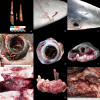
The panel displays the fragments extracted from the various blue sharks described in this article. Each figure is labeled with the number corresponding to the case to which the rostra belong. Most of the images include a metric reference (either a ruler or a grid with 1 cm squares). In those cases where such a reference was not originally present, a 1 cm scale bar has been added. In Case 1, the fragment was an incidental finding observed in the eye—previously fixed in formalin—during histological processing. In Case 2, no fragments were extracted, as the swordfish rostrum entered and exited the animal, leaving only a wound. Case 4 contained three fragments: two are shown on the photographic tray (4a) and the third within the animal itself (4b). All remaining cases show the complete set of fragments extracted from each shark.
Fig. 4
Fig. 4
Gross lesions from interspecific interactions between blue sharks and swordfish. (a) Five fragments from three different rostra in case 9. (b) Flattened ulcer with two tips from a hard and brownish foreign body coming out from opposite edges of the lesion. Case 7. (c) A subtle lesion in the skin that is cranio-dorsal to the right eye and resembling a minor superficial wound. Case 6. (d) Ulcerative scleritis with ocular perforation and the presence of hyphema. Case (1) (e) Flattened corneal perforation in the centre of the eye. Case (2) (f) Deep lesion with a penetrating rostrum through the left eye and into chondrocranium. Case 4. (g) Meningeal hemorrhage in response to a rostrum wound. Case 7. (h) Encephalon completely pierced by a rostrum fragment. Case 8. (i) Fibrous branchitis in response to the rostrum wound through the gill arches. Case 9.
Fig. 5
Fig. 5
Histological lesions from interspecific interactions between blue sharks and swordfish. (a) Abundant hemorrhage surrounding a rostrum wound. Case 6. Hematoxylin and eosin (HE). (b) Area of acute inflammation characterized by hemorrhage, fibrin, and mainly heterophilic infiltrate. Case 4. HE. (c) Eye with severe hemorrhage and fibrin. Case 2. HE. (d) Eye (lens). Severe cataract with presence of Morgagnian globules. Case 1. HE. (e) Severe hemorrhage in the brain caused by a rostrum. Case 6. HE. (f) Severe meningitis with presence of perivascular cuffings. Case 7. HE. (g) Chronic lesion characterized by dense connective tissue, with numerous collagen fibers (which stain blue) replacing the previously existing tissue skeletal muscle. Case 9. Masson’s trichrome. (h) Severe fibrosis in the gills associated with a chronic wound caused by a rostrum. Case 9. Masson’s trichrome. (i) Membranoproliferative glomerulonephritis. Case 4. HE. Inset: normal glomerulus from Case 9. HE.

Similar articles

References

    1. Smith, J. Pugnacity of Marlins and swordfish. Nature178, (1956).
    1. Ellis, R. Armed and angerous. in Swordfish: A Biography of the Ocean Gladiator 61–90 (University of Chicago Press, Chicago, (2013).
    1. Romeo, T., Amendola, G., Canese, S., Andaloro, F. & Battaglia, P. Recent records of swordfish attacks on harpoon vessels in the Sicilian waters (Mediterranean Sea). Acta Adriat (Online). 58, 147–156 (2017).
    1. Penadés-Suay, J., García-Salinas, P., Tomás, J. & Aznar, F. J. Aggressive interactions between juvenile swordfishes and blue sharks in the Western mediterranean: a widespread phenomenon? Medit Mar. Sci.20, 314–319 (2019).
    1. Machida, S. A swordfish sword found from a North Pacific Sei Whale. Sci. Rep. Whales Res. Inst.22, 163–164 (1970).

LinkOut - more resources