. 2025 Aug 12;13(1):72.
doi: 10.1038/s41413-025-00449-6.

The mitochondrial E3 ligase MAPL SUMOylates Drp1 to facilitate mitochondrial fission in intervertebral disc degeneration

Affiliations

The mitochondrial E3 ligase MAPL SUMOylates Drp1 to facilitate mitochondrial fission in intervertebral disc degeneration

Zhidi Lin et al. Bone Res. .

Abstract

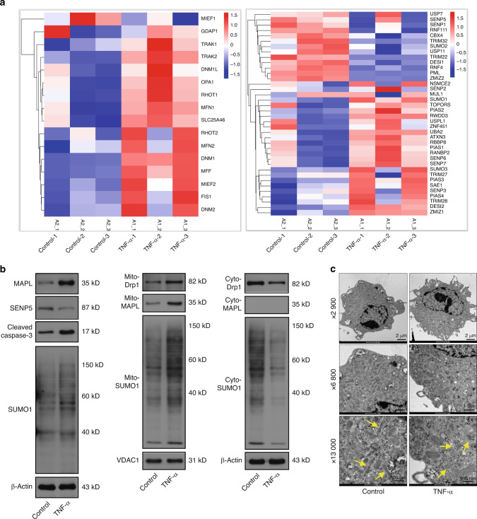
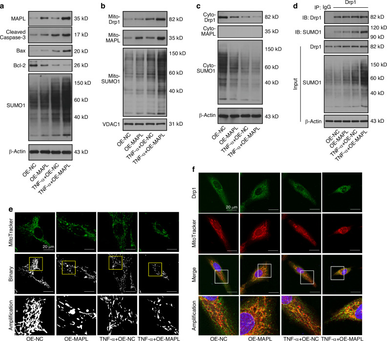
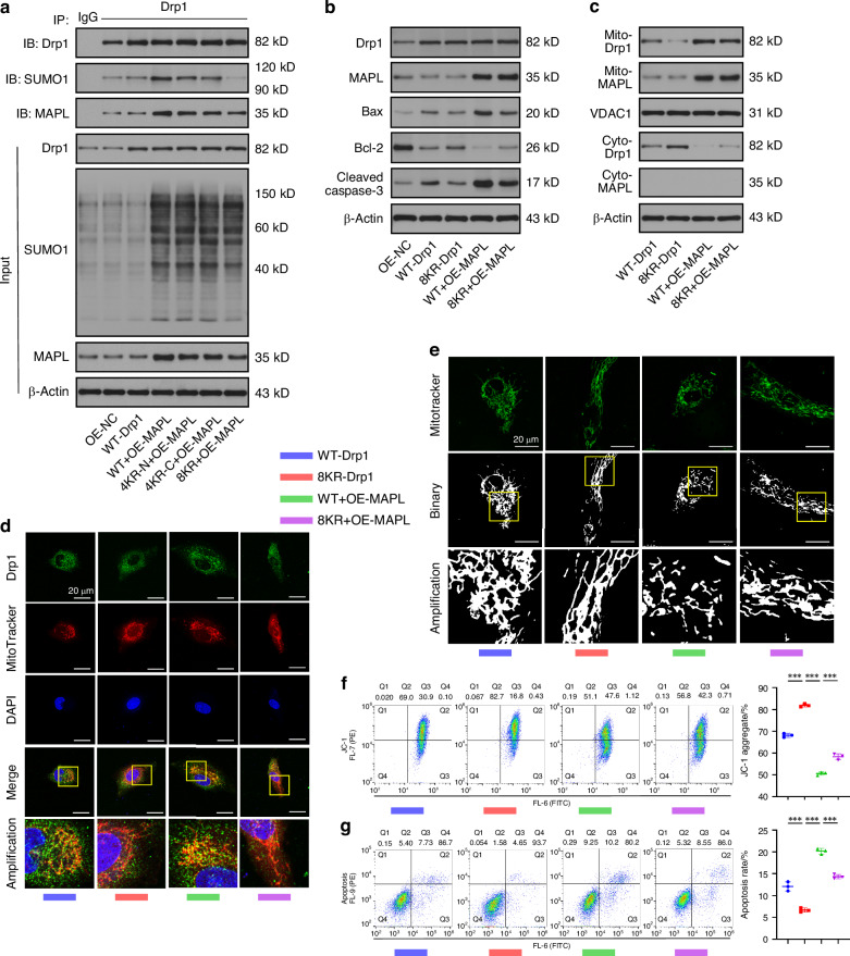
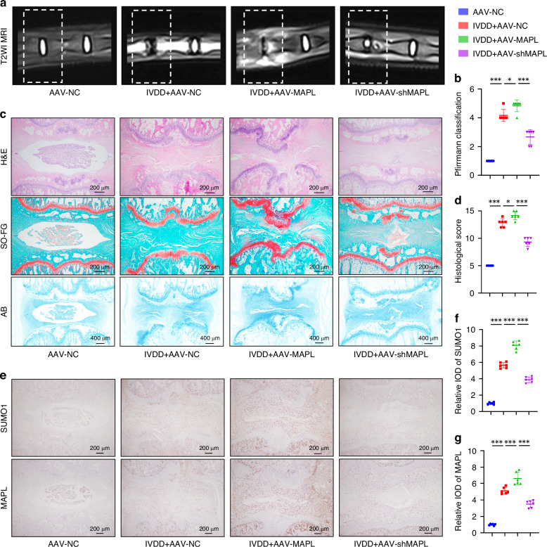
Intervertebral disc degeneration (IVDD) is the primary contributor to a range of spinal diseases. Dynamin-related protein 1 (Drp1)-mediated mitochondrial fission has recently been identified as a new cause of nucleus pulposus cell (NPC) death and IVDD, but the underlying mechanisms remain unclear. Although the effects of Drp1 phosphorylation in IVDD have been studied, it is currently unknown if small ubiquitin-like modifications (SUMOylation) of Drp1 regulate IVDD. This study aimed to investigate the functions and mechanisms of mitochondria-anchored protein ligase (MAPL), a mitochondrial SUMO E3 ligase, during IVDD progression. The expression of genes related to SUMOylation and mitochondrial dynamics in TNF-α-stimulated NPCs was analysed via RNA sequencing. The levels of total and mitochondrial SUMO1 conjugates were elevated with MAPL upregulation in TNF-α-treated NPCs. Additionally, mitochondrial fragmentation and dysfunction were induced by TNF-α stimulation. MAPL overexpression promoted mitochondrial SUMOylation and SUMO1 modification of Drp1, thereby facilitating the mitochondrial translocation of Drp1 and mitochondrial fission. MAPL-induced ROS accumulation and ΔΨm loss led to increased NPC apoptosis. Mutation of the SUMO-acceptor lysine residues of Drp1 hindered its SUMOylation and rescued the mitochondrial phenotypes caused by MAPL. SENP5 overexpression phenocopied MAPL silencing, negatively modulating the SUMO1 modification of Drp1 and mitochondrial fission in NPCs. In a rat IVDD model, forced expression of MAPL by using an adeno-associated virus (AAV) vector aggravated IVD tissue damage, whereas the knockdown of MAPL delayed IVDD progression. Our findings highlight the importance of SUMOylation in IVDD. The inhibition of MAPL-mediated Drp1 SUMOylation alleviates mitochondrial fission and limits IVDD development, providing a potential strategy for IVDD treatment.


Conflict of interest statement

Competing interests: The authors declare no competing interests.

Figures

Fig. 1
Fig. 1
SUMOylation and mitochondrial dynamics are associated with NPC apoptosis. a RNA sequencing of NPCs treated with or without TNF-α to analyse the expression of SUMOylation-related genes and mitochondrial dynamics-related genes. b Western blot analysis of total MAPL, SENP5, cleaved caspase-3 and SUMO1 levels in TNF-α-treated NPCs. The mitochondrial and cytosolic levels of Drp1, MAPL and SUMO1 were also evaluated. c The morphological ultrastructural appearance of mitochondria in NPCs was observed via TEM. (n = 3; *P < 0.05, **P < 0.01, and ***P < 0.001)
Fig. 2
Fig. 2
MAPL SUMOylates Drp1 and facilitates its recruitment to mitochondria. a The levels of total MAPL, cleaved caspase-3, Bax, Bcl-2 and SUMO1 in TNF-α-stimulated NPCs transfected with the MAPL overexpression plasmid were analysed by Western blotting. b, c Western blot analysis of the mitochondrial and cytosolic levels of Drp1, MAPL, and SUMO1 in NPCs after MAPL overexpression with or without TNF-α treatment. d SUMO1 modification of Drp1 was analysed by co-IP in MAPL-overexpressing NPCs under inflammatory conditions. e Mitochondrial fragmentation caused by MAPL overexpression was observed via MitoTracker Green staining. f The colocalization of Drp1 and MitoTracker Red was observed via confocal microscopy. (n = 3; *P < 0.05, **P < 0.01, and ***P < 0.001)
Fig. 3
Fig. 3
MAPL knockdown restricts Drp1 SUMOylation and mitochondrial fragmentation. a Western blots showing the levels of MAPL, Bax, Bcl-2, cleaved caspase-3 and SUMO1 in TNF-α-treated NPCs transfected with si-MAPL. b, c Western blots showing the levels of Drp1, MAPL and SUMO1 in the cytosolic fraction and mitochondrial compartment of NPCs after MAPL knockdown. d Co-IP of SUMOylated Drp1 in NPCs after treatment with si-MAPL. e Mitochondrial ROS production in NPCs transfected with si-MAPL. f, g JC-1 staining and flow cytometry were used to assess the ΔΨm in NPCs after MAPL knockdown. h, i DCFH-DA staining and flow cytometry were used to detect ROS accumulation in NPCs. j, k Flow cytometry with Annexin V-FITC/PI staining was used to evaluate the apoptosis rate of NPCs transfected with si-MAPL. (n = 3; *P < 0.05, **P < 0.01, and ***P < 0.001)
Fig. 4
Fig. 4
Mutation of key lysine residues blocks Drp1 SUMOylation and rescues mitochondria-mediated NPC apoptosis. a MAPL overexpression-mediated SUMOylation of Drp1 was analysed via co-IP in NPCs transfected with WT-Drp1 or the 4KR-N, 4KR-C and 8KR mutants. b Western blots showing the levels of total Drp1, MAPL, cleaved caspase-3, Bax, and Bcl-2 in NPCs transfected with the WT-Drp1 or 8KR plasmid after MAPL was overexpressed. c Western blot analysis of Drp1 and MAPL levels in the cytosolic fraction and mitochondrial compartment of NPCs after MAPL overexpression and lysine mutation of Drp1. d The translocation of Drp1 in NPCs was observed using MitoTracker Red staining and immunofluorescence staining for Drp1. e Changes in the morphology of mitochondria in MAPL-overexpressing NPCs transfected with WT-Drp1 or 8KR, as shown by MitoTracker Green staining. f JC-1 staining and flow cytometry were used to detect the ΔΨm in NPCs transfected with WT-Drp1 or 8KR after MAPL overexpression. g Flow cytometry with Annexin V-FITC/PI staining was used to detect the apoptotic rate of NPCs. (n = 3; *P < 0.05, **P < 0.01, and ***P < 0.001)
Fig. 5
Fig. 5
SENP5 negatively modulates Drp1 SUMOylation and mitochondrial fission. a Western blots showing the levels of total SENP5, cleaved caspase-3 and SUMO1 in NPCs after transfection with the SENP5 overexpression plasmid under inflammatory conditions. b, c Drp1 and SUMO1 expression in the cytosolic and mitochondrial fractions of the TNF-α-treated NPCs after SENP5 overexpression was measured via Western blotting. d Co-IP analysis of the SENP5-induced SUMO1 modification of Drp1 after TNF-α treatment. e Western blot analysis of total SENP5, cleaved caspase-3 and SUMO1 levels in NPCs after SENP5 knockdown and TNF-α treatment. f, g Mitochondrial and cytosolic levels of Drp1 and SUMO1 in TNF-α-treated NPCs transfected with si-SENP5 were assessed by Western blotting. h Drp1 SUMOylation after SENP5 silencing was analysed via co-IP. (n = 3; *P < 0.05, **P < 0.01, and ***P < 0.001)
Fig. 6
Fig. 6
SUMOylation and mitochondrial translocation of Drp1 are coregulated by MAPL and SENP5. a Western blots showing the levels of total MAPL, SENP5, cleaved caspase-3, and SUMO1 in NPCs transfected with OE-MAPL or OE-SENP5 plasmids. b, c Mitochondrial and cytosolic levels of Drp1 and SUMO1 in MAPL-overexpressing and SENP5-overexpressing NPCs were assessed using Western blotting. d Co-IP of SUMOylated Drp1 in NPCs overexpressing MAPL and SENP. e MitoTracker Red staining and immunofluorescence staining for Drp1 in NPCs after MAPL or SENP5 overexpression. f Changes in the morphology of mitochondria in NPCs transfected with OE-MAPL or OE-SENP5, as shown by MitoTracker Green staining. (n = 3; *P < 0.05, **P < 0.01, and ***P < 0.001)
Fig. 7
Fig. 7
Inhibition of MAPL delays IVDD in a rat model. a, b The coccygeal vertebrae of the rats were examined by MRI, and the degree of IVDD was evaluated by using the Pfirrmann classification. c H&E staining, Safranin O‒Fast Green staining and Alcian blue staining of rat IVD samples. d Evaluation of IVDD by histological scoring. eg Immunohistochemical analysis of SUMO1 and MAPL levels. (n = 6; *P < 0.05, **P < 0.01, and ***P < 0.001)

References

    1. Mohd Isa, I. L. et al. Intervertebral disc degeneration: biomaterials and tissue engineering strategies toward precision medicine. Adv. Healthcare Mater.11, 2102530 (2022). - PMC - PubMed
    1. Qi, M. et al. Comparison of clinical outcomes between cervical disc arthroplasty and anterior cervical discectomy and fusion for the treatment of single-level cervical spondylosis: a 10-year follow-up study. Spine J.23, 361–368 (2023). - PubMed
    1. Wang, Z. et al. Inhibition of aberrant Hif1α activation delays intervertebral disc degeneration in adult mice. Bone Res.10, 2 (2022). - PMC - PubMed
    1. Chen, Z. et al. N6-methyladenosine hypomethylation of circGPATCH2L regulates DNA damage and apoptosis through TRIM28 in intervertebral disc degeneration. Cell Death Differ.30, 1957–1972 (2023). - PMC - PubMed
    1. Song, Y. et al. Mitochondrial quality control in intervertebral disc degeneration. Exp. Mol. Med.53, 1124–1133 (2021). - PMC - PubMed

MeSH terms

LinkOut - more resources