. 2025 Aug 12;16(1):7457.
doi: 10.1038/s41467-025-62399-1.

COUP-TFII-mediated reprogramming of the vascular endothelium counteracts tumor immune evasion

Affiliations

COUP-TFII-mediated reprogramming of the vascular endothelium counteracts tumor immune evasion

Yu Zhu et al. Nat Commun. .

Abstract

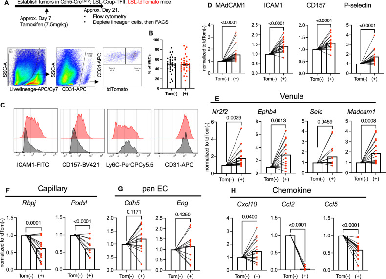
T cell scarcity in tumor tissues poses a critical challenge to cancer immunotherapy. Here we manipulate the tumor vasculature, an essential regulator of immune cell trafficking, to reinvigorate anti-tumor T cell responses in "cold" tumors. We show that ectopic pan-endothelial expression of COUP-TFII, a master transcription factor for venous development, induces molecular programs of post-capillary venules in tumor endothelium. Venular reprogramming selectively promotes T cell recruitment into tumors, inhibits tumor growth in mouse models of breast and pancreatic cancers, and sensitizes tumors to immune checkpoint blockade and adoptive T cell transfer therapies. Mechanistic studies show that enhanced recruitment of anti-tumor T cells and tumor inhibition are mediated by COUP-TFII-induced vascular adhesion receptors. Our study supports a pivotal role of vascular endothelial cells in governing tumor immune evasion, and proposes venular reprogramming as a therapeutic strategy to bolster anti-tumor immunity and immunotherapy.


Conflict of interest statement

Competing interests: The authors declare no competing interests.

Figures

Fig. 1
Fig. 1. Tumor expansion downregulates T cell infiltration and venular endothelial specialization.
A Representative flow cytometry gating strategy for vascular blood endothelial cells (BEC) in MMTV-PyMT tumors. B, C Flow cytometric quantification of tumor-associated BEC abundance. DF Quantification of mean fluorescence intensity (MFI) of indicated markers in tumor BECs. GN Flow cytometric quantification of the frequencies of indicated leukocytes in autochthonous MMTV-PyMT-B6 tumors from 4-month-old mice. BN small: n = 9 biological replicates; large: n = 11 biological replicates). Representative of two independent experiments. Groups were compared by an unpaired two-tailed Student’s t-test. Error bars indicate s.e.m.
Fig. 2
Fig. 2. Ectopic expression of COUP-TFII in endothelial cells drives capillary-to-venule reprogramming.
A, B Representative histogram and MFI quantification of COUP-TFII in BECs from orthotopic PyMT tumors established in iCoup and control mice. Pre-gated on BECs. Representative of three independent experiments. (n = 7 biological replicates/group). C Representative immunofluorescence imaging of COUP-TFII and CD31 in orthotopic PyMT tumors established in iCoup and control mice. Scale bar represents 50 μm. D Flow cytometry quantification of frequency of P-selectin+ venular BECs. EL MFI of indicated markers in BECs from orthotopic PyMT tumors established in iCoup and control mice (DF, IK control: n = 7 biological replicates, iCoup: n = 5 biological replicates; G, H, Ln = 5 biological replicates/group). MO Representative histograms and contour plots of indicated molecules in BECs from orthotopic PyMT tumors of iCoup and control mice. Pre-gated on BECs. P MFI of F2 antibody staining in BECs from orthotopic PyMT tumors established in iCoup and control mice (Control: n = 7 biological replicates, iCoup: n = 5 biological replicates). Q Representative histograms of PNAd in BECs from orthotopic PyMT tumors of iCoup and control mice. Pre-gated on BECs. Data in (DN) are representative of more than five independent experiments. Data in (OQ) are representative of three independent experiments. Groups were compared by unpaired two-tailed Student’s t-test. Error bars indicate s.e.m.
Fig. 3
Fig. 3. Ectopic expression of COUP-TFII reprograms ECs in a cell-autonomous manner.
A Schematics of mosaic COUP-TFII induction in tumor BECs and gating strategy. Lineage+ (Ter119+/EpCAM+/CD45+/Podoplanin+) cells were depleted from digested tumors before proceeding to fluorescence-activated cell sorting (FACS). B Quantification of tdTomato (Tom+) labeling efficiency in (A). (n = 32/group). C, D Representative histograms (C) and MFI quantification (D) of indicated marker expression in tdTomato(−) and (+) BECs from orthotopic tumors; MFI values of tdTom(+) BECs were normalized internally to those of the tdTom(−) counterpart within the same mouse, and pairwise comparison was performed (n = 25 biological replicates/group for MAdCAM1 and CD157, n = 23 biological replicates/group for ICAM1, n = 13 biological replicates/group for P-selectin). Representative of three independent repeats. EH QPCR analyses of indicated molecules of tdTomato(−) and (+) BECs sorted from orthotopic tumors; gene expression values from each mouse were normalized internally to that of the tdTomato(−) BECs and pairwise comparison was performed (n = 19 biological replicates/group for Nr2f2 and Madcam1; n = 16 biological replicates/group for Ephb4, Sele and Ccl5; n = 15 biological replicates/group for Rbpj and Cxcl10; n = 12 biological replicates/group for Eng, n = 11 biological replicates/group for Podxl and Ccl2; n = 14 biological replicates/group for Cdh5). Groups were compared by unpaired two-tailed Student’s t-test. Error bars indicate s.e.m.
Fig. 4
Fig. 4. COUP-TFII-driven capillary-to-venule reprogramming enhances T cell infiltration.
AC Flow cytometric quantification of the frequency of indicated lymphocytes among cells dissociated from orthotopic PyMT-B6 tumors in iCoup and control mice, based on gating strategies in Supplementary Fig. 2; representative of three independent experiments. (n = 6 biological replicates/group). DK Flow cytometric quantification of the frequency of indicated leukocytes among cells dissociated from orthotopic PyMT-B6 tumors in iCoup and control mice, based on gating strategies in Supplementary Fig. 2; representative of three independent experiments. (n = 5 biological replicates/group). L, M Representative immunofluorescence imaging (L) and quantification (M) of CD8+ cells in GEMM tumors from 5-month-old MMTV-PyMT-iCoup and control MMTV-PyMT mice. Scale bars represent 100 μm. (n = 5 biological replicates/group). N Representative immunofluorescence imaging of CD8, CD31, and keratin of orthotopic KPC tumors established in control and iCoup mice. Scale bars represent 100 μm. O, P Flow cytometric quantification of BrdU incorporation in tumor-infiltrating CD8+ and CD4 + T cells. Representative of two independent experiments. (n = 6 biological replicates/group). Indicated cell populations are quantified as % of total cells dissociated from the tumor, based on gating strategies from Supplementary Fig. 2. Groups were compared by unpaired two-tailed Student’s t-test. Error bars indicate s.e.m.
Fig. 5
Fig. 5. COUP-TFII-driven capillary-to-venule reprogramming enhances T cell recruitment from circulation.
A Schematics and quantification of lymph node T cell homing assay in PyMT-B6 tumors. (n = 8 biological replicates/group). Representative of more than three independent experiments. B Schematics, representative flow cytometry plots, and quantification of OT1 T cell homing to orthotopic PyMT-OVA tumors. (n = 3 biological replicates/group). Representative of three independent experiments. C Flow cytometric analyses of bone marrow cell homing to orthotopic PyMT tumors. (n = 8 biological replicates/group). Representative of more than three independent experiments. Quantification in (B, C) indicates the frequency of homed donor-derived leukocytes as % of total cells dissociated from the tumor. D Gating strategy for circulating leukocytes in the blood. E Frequencies of circulating leukocyte subsets in the blood of iCoup and control mice, quantified as the percentage among circulating CD45+ cells. (n = 4 biological replicates/group). FI Frequencies of indicated T cell populations in the tumor-draining lymph nodes (dLNs). (control: n = 10 biological replicates, iCoup: n = 12 biological replicates). JQ Representative gating strategy and flow cytometric quantifications of indicated T cell subsets in the dLNs. (control: n = 10 biological replicates, iCoup n = 12 biological replicates). Groups were compared by an unpaired two-tailed Student’s t-test, ns denotes not significant. Error bars indicate s.e.m.
Fig. 6
Fig. 6. COUP-TFII-driven endothelial reprogramming inhibits tumor growth in a T cell-dependent manner.
AC Tumor volume or wet weight measurements of subcutaneous (subQ) (A), orthotopic (B), or autochthonous (C) PyMT tumors in iCoup and control mice. A, B is representative of more than five independent experiments. (A control: n = 6, iCoup: n = 7; B control: n = 5, iCoup: n = 3; C control: n = 9, iCoup: n = 16). D Survival curve of MMTV-PyMT-iCoup and control MMTV-PyMT mice. ****p < 0.0001 by log-rank (Mantel–Cox) test. (control: n = 17, iCoup: n = 13). E, F Tumor volume and wet weight measurements of orthotopic PyMT tumors in iCoup and control mice treated with CD8-depleting antibodies or control IgGs. (control IgG: n = 3, control αCD8: n = 4, iCoup IgG: n = 6, iCoup αCD8: n = 5). G, H Tumor volume or wet weight measurements of subcutaneous (A) and orthotopic (B) KPC tumors in iCoup and control mice. (n = 3–4/group). (G control: n = 3, iCoup: n = 4; H n = 4/group). Representative of more than three independent experiments. I Wet weight measurements of orthotopic KPC tumors in iCoup and control mice treated with CD8-depleting antibodies. (control IgG: n = 6, control αCD8: n = 5, iCoup IgG: n = 6, iCoup αCD8: n = 5). Groups were compared by two-way ANOVA (A, E), unpaired two-tailed Student’s t-test (B, C, FI), or log-rank (Mantel–Cox) test (D). Error bars indicate s.e.m.
Fig. 7
Fig. 7. Venule-mediated antitumor T cell recruitment requires COUP-TFII induction of E/P-selectins and CXCR3 signaling.
A, B Flow cytometric quantification of the frequency of tumor-infiltrating CD4+ or CD8 + T cells in subcutaneous PyMT-bearing iCoup or control mice, treated with E- and P-selectin blocking antibodies or control IgGs. C tumor volume measurement of mice in (A, B). AC Control IgG: n = 5, Control αE/P-selectin: n = 5, iCoup IgG: n = 6, iCoup αE/P-selectin: n = 5. Representative of two independent experiments. D, E Flow cytometric quantification of tumor-infiltrating CD4+ and CD8 + T cells in subcutaneous PyMT-OVA-bearing iCoup or control mice, treated with CXCR3 blocking antibodies or control IgGs. (n = 6–7/group). F Tumor volume measurement of mice in (D, E). DF Control IgG: n = 7, Control αCXCR3: n = 6, iCoup IgG: n = 7, iCoup αCXCR3: n = 6. Representative of three independent experiments. Groups were compared by unpaired two-tailed Student’s t-test (A, B, D, E) or two-way ANOVA (C, F). Error bars indicate s.e.m.
Fig. 8
Fig. 8. COUP-TFII-driven venule programming sensitizes PyMT and KPC tumor responses to immunotherapies.
A, B Wet weight measurements of subcutaneous PyMT (A) and subcutaneous KPC tumors (B) in iCoup and control mice treated with αCTLA4 and αPD1 or control antibodies. (A control IgG: n = 6, control αPD1/CTLA4: n = 4, iCoup IgG: n = 6, iCoup αPD1/CTLA4: n = 5; B control IgG: n = 4, control αPD1/CTLA4: n = 3, iCoup IgG: n = 3, iCoup αPD1/CTLA4: n = 4). C, D Wet weight measurements of orthotopic PyMT-OVA (C) and KPC-OVA (D) tumors in iCoup and control mice treated with ex vivo-activated OT1 cells. (C control: n = 4, control OT1: n = 5, iCoup: n = 5, iCoup OT1: n = 5; B control: n = 4, control OT1: n = 3, iCoup: n = 3, iCoup OT1: n = 4). Groups were compared by unpaired two-tailed Student’s t-test unless. Error bars indicate s.e.m. Representative of two independent experiments. n = 3–6/group.

References

    1. Waldman, A. D., Fritz, J. M. & Lenardo, M. J. A guide to cancer immunotherapy: from T cell basic science to clinical practice. Nat. Rev. Immunol.20, 651–668 (2020). - PMC - PubMed
    1. Sharma, P., Hu-Lieskovan, S., Wargo, J. A. & Ribas, A. Primary, adaptive, and acquired resistance to cancer immunotherapy. Cell168, 707–723 (2017). - PMC - PubMed
    1. Morrison, A. H., Byrne, K. T. & Vonderheide, R. H. Immunotherapy and prevention of pancreatic cancer. Trends Cancer4, 418–428 (2018). - PMC - PubMed
    1. Ochoa de Olza, M., Navarro Rodrigo, B., Zimmermann, S. & Coukos, G. Turning up the heat on non-immunoreactive tumours: opportunities for clinical development. Lancet Oncol.21, e419–e430 (2020). - PubMed
    1. Joyce, J. A. & Fearon, D. T. T cell exclusion, immune privilege, and the tumor microenvironment. Science348, 74–80 (2015). - PubMed

MeSH terms

Substances